Copyright
©The Author(s) 2025.
World J Gastroenterol. May 14, 2025; 31(18): 105729
Published online May 14, 2025. doi: 10.3748/wjg.v31.i18.105729
Published online May 14, 2025. doi: 10.3748/wjg.v31.i18.105729
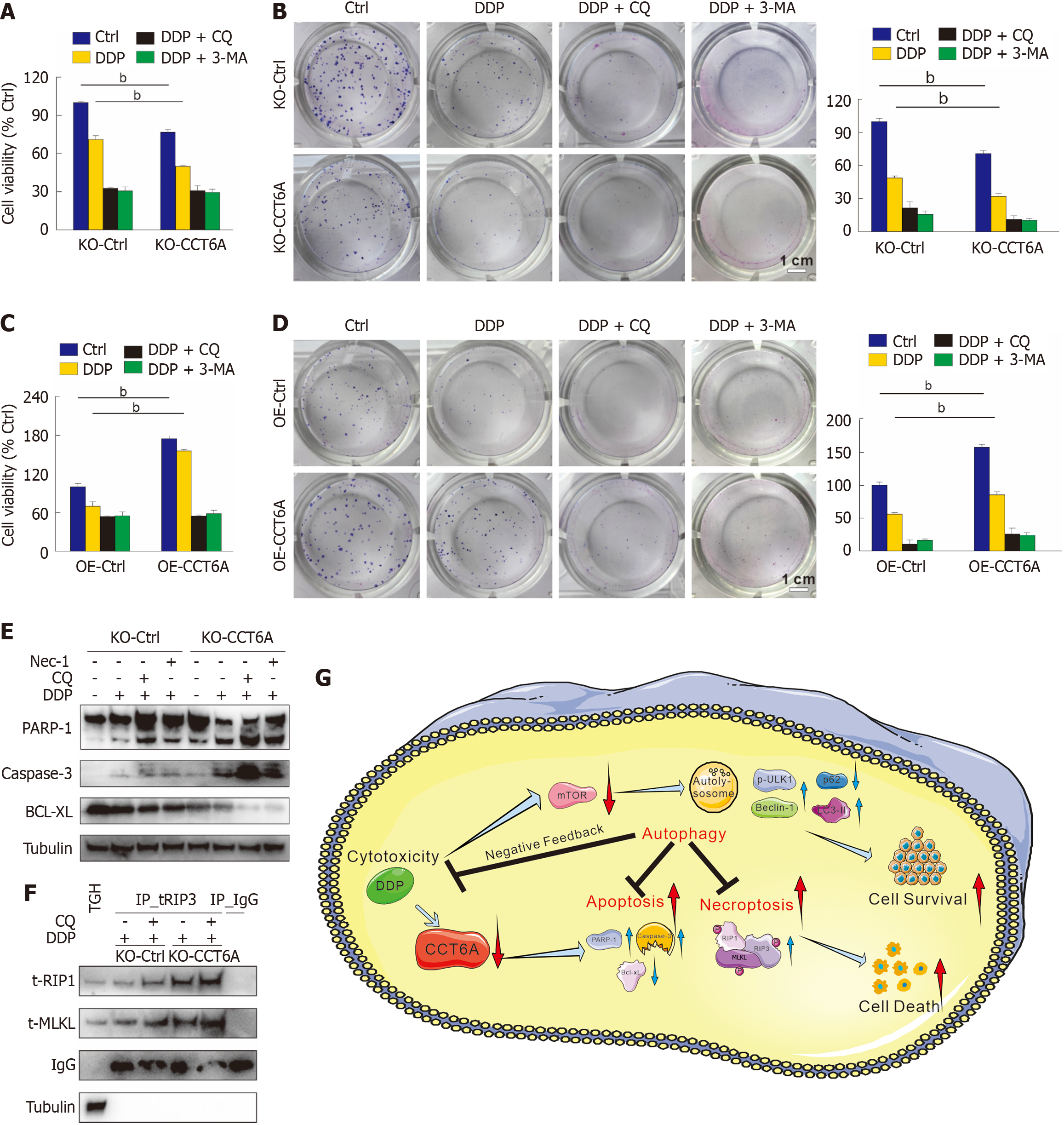
Figure 7 Inhibition of autophagy enhances the cytotoxicity of cisplatin.
A-D: The MTS assay was conducted to assess the cell viability after applying the specified treatments for 24 hours (3-Methyladenine [3-MA]: 2 mM) (A and C), colony growth assay was performed with indicated treatments (cisplatin [DDP]: 1 μM; chloroquine diphosphate salt [CQ]: 5 μM; 3-MA: 0.5 mmol/L) (scale bar = 1 cm) (B and D); E: Following appropriate treatments for 24 hours, cell lysates were performed immunoblotting with indicated antibodies; F: Co-immunoblotting assay was conducted with the receptor-interacting protein kinase 3 antibody, and then subjected to immunoblotting; G: Schematic model of chaperonin-containing tailless complex polypeptide 1 subunit 6a (CCT6A) modulates the cytotoxicity of DDP via regulating autophagy. bP < 0.01 vs control (Ctrl). Bcl-xL: B-cell lymphoma-extra large; IgG: Immunoglobulin G (negative control antibody); MLKL: Mixed lineage kinase domain-like; Nec-1; Necrostatin-1; PARP-1; Poly(ADP-ribose) polymerase 1; RIP1: Receptor-interacting protein kinase 1; TH: Total homogenate.
- Citation: Ma JX, Li XJ, Li YL, Liu MC, Du RH, Cheng Y, Li LJ, Ai ZY, Jiang JT, Yan SY. Chaperonin-containing tailless complex polypeptide 1 subunit 6A negatively regulates autophagy and protects colorectal cancer cells from cisplatin-induced cytotoxicity. World J Gastroenterol 2025; 31(18): 105729
- URL: https://www.wjgnet.com/1007-9327/full/v31/i18/105729.htm
- DOI: https://dx.doi.org/10.3748/wjg.v31.i18.105729


