Copyright
©The Author(s) 2025.
World J Gastroenterol. May 14, 2025; 31(18): 105729
Published online May 14, 2025. doi: 10.3748/wjg.v31.i18.105729
Published online May 14, 2025. doi: 10.3748/wjg.v31.i18.105729
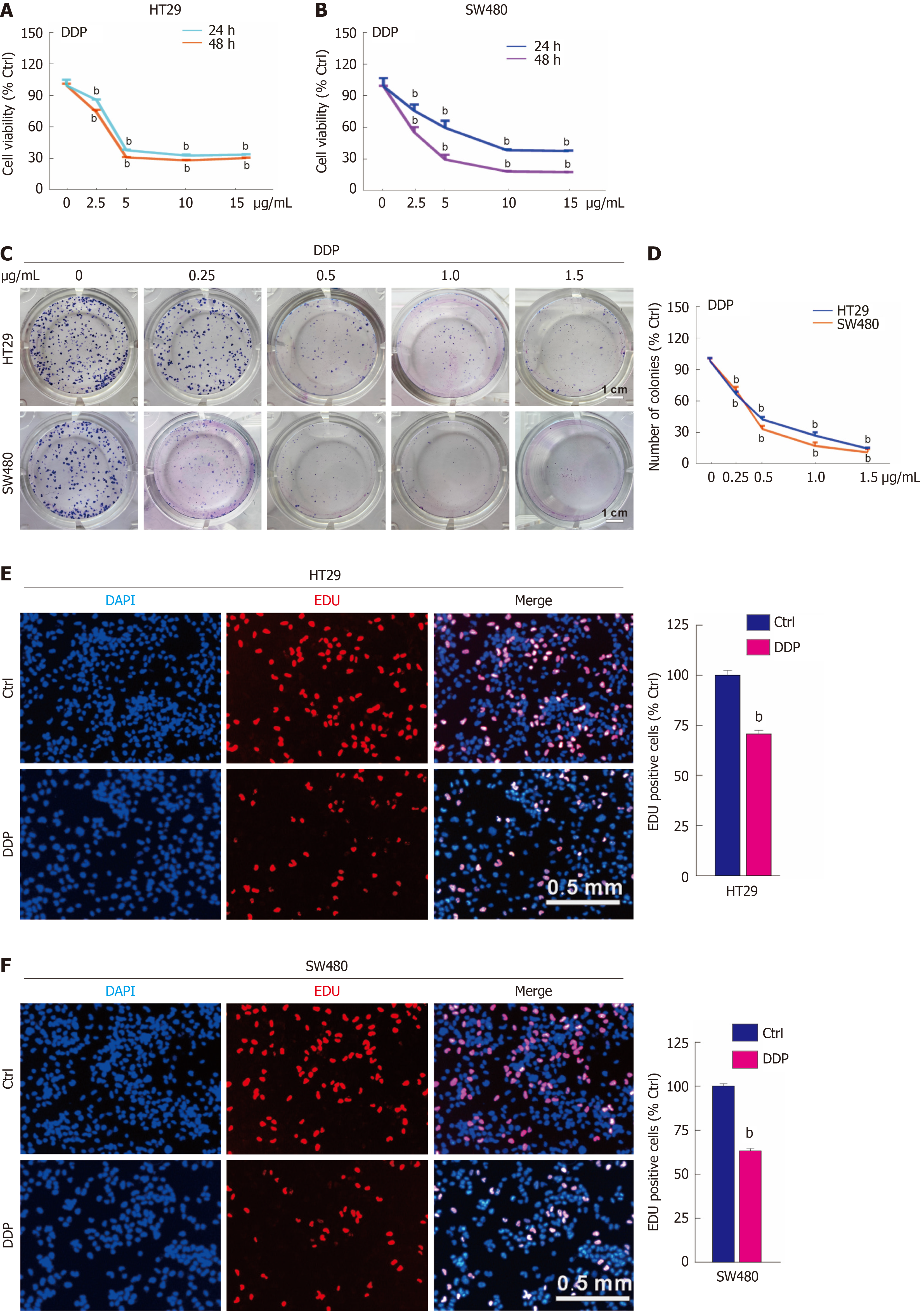
Figure 1 Cisplatin reduces cell proliferation in colorectal cancer.
A and B: Indicated concentrations of cisplatin (DDP) (μg/mL) were used to treat HT29 and SW480 cells, and the cell viability was measured by MTS assay; C and D: For the colony growth assay, HT29 and SW480 were treated with the indicated concentration gradient of DDP (μg/mL), and colony formation numbers were calculated and graphically represented using ImageJ and Prism GraphPad software. Scale bar = 1 cm; E and F: EdU staining assay was conducted to evaluate cell proliferation following DDP treatment (5 μg/mL, hereafter unless otherwise indicated) for 3 hours. Scale bar = 0.5 mm. bP < 0.01 vs control (Ctrl).
- Citation: Ma JX, Li XJ, Li YL, Liu MC, Du RH, Cheng Y, Li LJ, Ai ZY, Jiang JT, Yan SY. Chaperonin-containing tailless complex polypeptide 1 subunit 6A negatively regulates autophagy and protects colorectal cancer cells from cisplatin-induced cytotoxicity. World J Gastroenterol 2025; 31(18): 105729
- URL: https://www.wjgnet.com/1007-9327/full/v31/i18/105729.htm
- DOI: https://dx.doi.org/10.3748/wjg.v31.i18.105729


