Copyright
©The Author(s) 2025.
World J Gastroenterol. Apr 14, 2025; 31(14): 104975
Published online Apr 14, 2025. doi: 10.3748/wjg.v31.i14.104975
Published online Apr 14, 2025. doi: 10.3748/wjg.v31.i14.104975
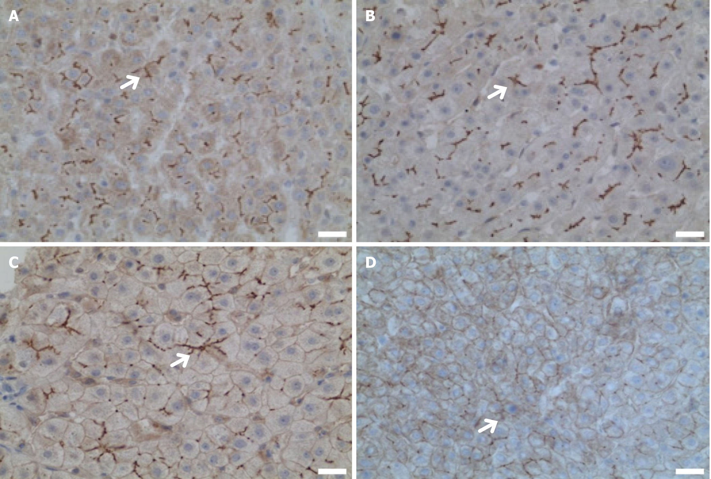
Figure 3 Immunohistochemical manifestations of the liver in a patient with ATP-binding cassette subfamily B member 4 variants.
Except for patient 4, all other patients underwent liver biopsy. Immunohistochemical staining was performed using anti- ATP-binding cassette subfamily B member 4 (P3II-26, Thermo, United States), with the white arrows indicating the labeling of multidrug resistance protein 3 (MDR3) tubules. Bars = 2.5 μm. A: MDR3 is evenly distributed in liver tissue (the white arrow indicates MDR3 is normal, case 1); B: MDR3 is evenly distributed in liver tissue (the white arrow indicates MDR3 is normal, 400 ×, case 2); C: Decreased distribution of MDR3 in liver tissue (the white arrow indicates MDR3 is decreasing, 400 ×, case 3); D: Decreased distribution of MDR3 in liver tissue (the white arrow indicates MDR3 is decreasing, 400 ×, case 5). ABCB4: ATP-binding cassette subfamily B member 4; MDR3: Multidrug resistance protein 3.
- Citation: Weng YH, Zheng YF, Yin DD, Xiong QF, Li JL, Li SX, Chen W, Yang YF. Clinical, genetic and functional perspectives on ATP-binding cassette subfamily B member 4 variants in five cholestasis adults. World J Gastroenterol 2025; 31(14): 104975
- URL: https://www.wjgnet.com/1007-9327/full/v31/i14/104975.htm
- DOI: https://dx.doi.org/10.3748/wjg.v31.i14.104975


