Copyright
©The Author(s) 2025.
World J Gastroenterol. Apr 14, 2025; 31(14): 104975
Published online Apr 14, 2025. doi: 10.3748/wjg.v31.i14.104975
Published online Apr 14, 2025. doi: 10.3748/wjg.v31.i14.104975
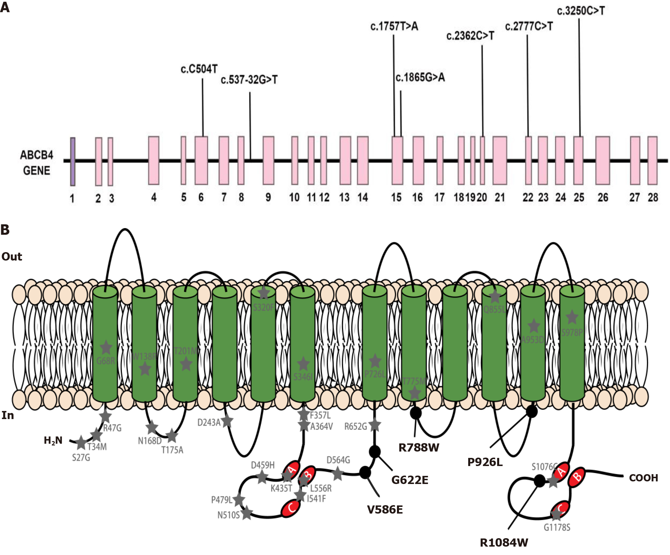
Figure 1 Schematic representation of ATP-binding cassette subfamily B member 4.
A: ATP-binding cassette subfamily B member 4 (ABCB4) gene structure with mutations representing exons 1 to 28. Each mutation found in our study is marked; B: The secondary structure of multidrug resistance protein 3 protein and the distribution of missense mutations found by our team. Black circle marked ABCB4 variants were detected in the patients cohort in our research. Grey pentagram marked ABCB4 variants reported in the literature; A: Walker A; B: Walker B; C: The signature motif C; COOH: Carboxyl terminus of polypeptide chain; H2N: Amino terminus of polypeptide chain; In: Inside the cell membrane; Out: Outside the cell membrane; ABCB4: ATP-binding cassette subfamily B member 4; MDR3: Multidrug resistance protein 3.
- Citation: Weng YH, Zheng YF, Yin DD, Xiong QF, Li JL, Li SX, Chen W, Yang YF. Clinical, genetic and functional perspectives on ATP-binding cassette subfamily B member 4 variants in five cholestasis adults. World J Gastroenterol 2025; 31(14): 104975
- URL: https://www.wjgnet.com/1007-9327/full/v31/i14/104975.htm
- DOI: https://dx.doi.org/10.3748/wjg.v31.i14.104975


