Copyright
©The Author(s) 2024.
World J Gastroenterol. Jan 28, 2024; 30(4): 367-380
Published online Jan 28, 2024. doi: 10.3748/wjg.v30.i4.367
Published online Jan 28, 2024. doi: 10.3748/wjg.v30.i4.367
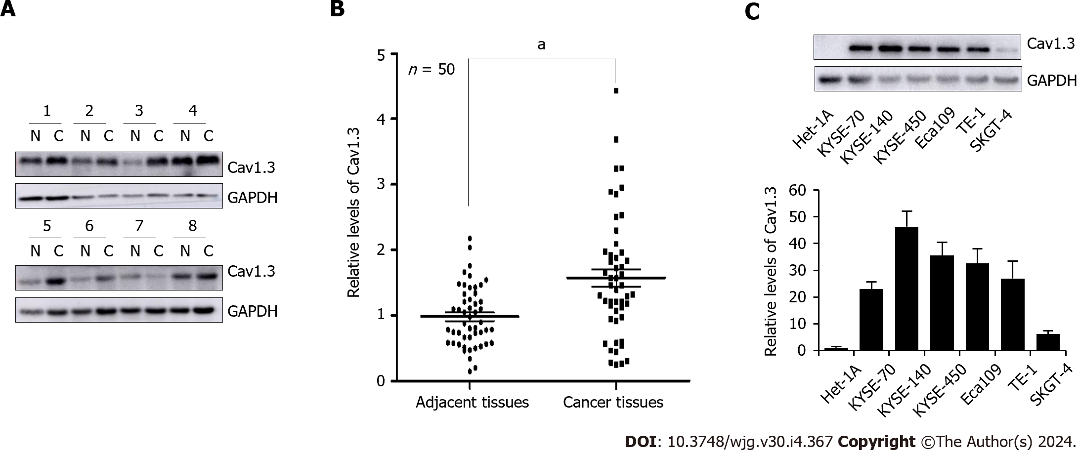
Figure 1 Levels of Cav1.
3 expression in esophageal cancer cell tissues. A: Representative detection of Cav1.3 level in esophageal squamous cell carcinoma (ESCC) by Western blot, N means adjacent tissues and C means cancer tissues; B: Statistical analysis of 50 cases of esophageal squamous cell carcinoma showed that level of Cav1.3 in ESCC tumor tissue was 1.60 times higher than that in adjacent tissue. aP < 0.05 vs the adjacent tissues; C: Western blot detection showed that the level of Cav1.3 in ESCC cells KYSE-70, KYSE-140, KYSE-450, Eca109, TE-1, and esophageal adenocarcinoma cell SKGT-4 was higher than that in esophageal squamous cell Het-1A.
- Citation: Chen YM, Yang WQ, Gu CW, Fan YY, Liu YZ, Zhao BS. Amlodipine inhibits the proliferation and migration of esophageal carcinoma cells through the induction of endoplasmic reticulum stress. World J Gastroenterol 2024; 30(4): 367-380
- URL: https://www.wjgnet.com/1007-9327/full/v30/i4/367.htm
- DOI: https://dx.doi.org/10.3748/wjg.v30.i4.367


