Copyright
©The Author(s) 2024.
World J Gastroenterol. May 7, 2024; 30(17): 2332-2342
Published online May 7, 2024. doi: 10.3748/wjg.v30.i17.2332
Published online May 7, 2024. doi: 10.3748/wjg.v30.i17.2332
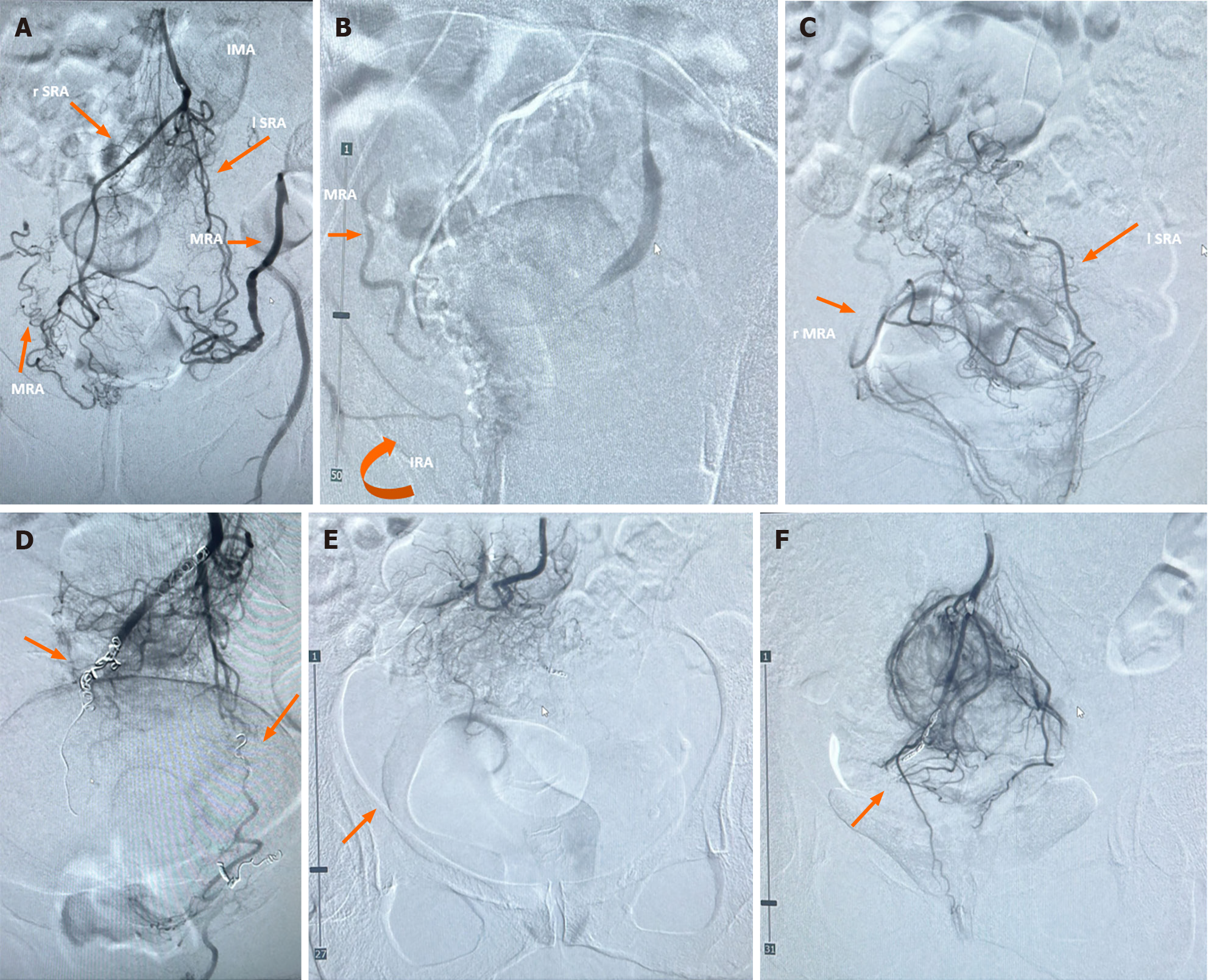
Figure 3 Pre-treatment and post-treatment angiography.
A: Case 1, anastomosis between superior (SRA) and middle (MRA) rectal arteries, bilaterally; B: Case 5, anastomosis between right MRA and inferior (IRA) rectal arteries; C: Case 8, anastomosis between right MRA and left SRA; D: Case 1, embolization of the distal branches of the SRA (arrows) and occlusion of the anastomosis with MRA; E: Case 5, embolization of the distal branches of the SRA and occlusion of the anastomosis with IRA and MRA (arrow); F: Case 8, embolization of the distal branches of the SRA (arrow) and occlusion of the anastomosis between right MRA and left SRA. IMA: Inferior mesenteric artery.
- Citation: Tutino R, Stecca T, Farneti F, Massani M, Santoro GA. Transanal eco-Doppler evaluation after hemorrhoidal artery embolization. World J Gastroenterol 2024; 30(17): 2332-2342
- URL: https://www.wjgnet.com/1007-9327/full/v30/i17/2332.htm
- DOI: https://dx.doi.org/10.3748/wjg.v30.i17.2332


