Copyright
©The Author(s) 2024.
World J Gastroenterol. Mar 21, 2024; 30(11): 1636-1643
Published online Mar 21, 2024. doi: 10.3748/wjg.v30.i11.1636
Published online Mar 21, 2024. doi: 10.3748/wjg.v30.i11.1636
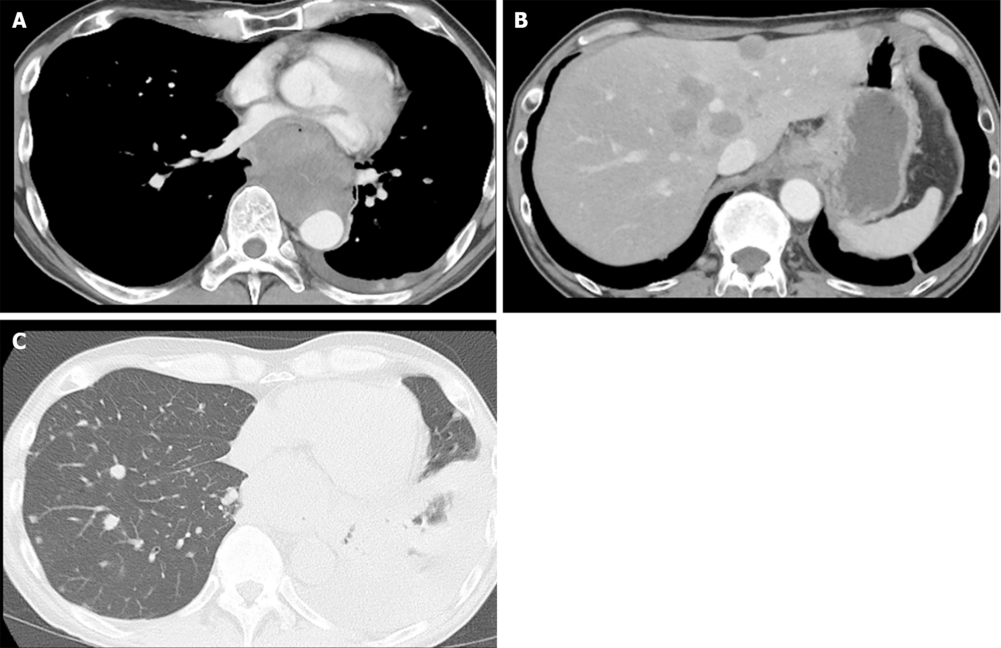
Figure 2 Imaging of primary and metastatic lesions by computed tomography.
A: Contrast-enhanced computed tomography shows left atrial compression due to the esophageal tumor; B: Multiple liver metastases; C: Multiple lung metastases and left pleural effusion.
- Citation: Shibata Y, Ohmura H, Komatsu K, Sagara K, Matsuyama A, Nakano R, Baba E. Myocardial metastasis from ZEB1- and TWIST-positive spindle cell carcinoma of the esophagus: A case report. World J Gastroenterol 2024; 30(11): 1636-1643
- URL: https://www.wjgnet.com/1007-9327/full/v30/i11/1636.htm
- DOI: https://dx.doi.org/10.3748/wjg.v30.i11.1636


