Copyright
©The Author(s) 2023.
World J Gastroenterol. Feb 21, 2023; 29(7): 1235-1242
Published online Feb 21, 2023. doi: 10.3748/wjg.v29.i7.1235
Published online Feb 21, 2023. doi: 10.3748/wjg.v29.i7.1235
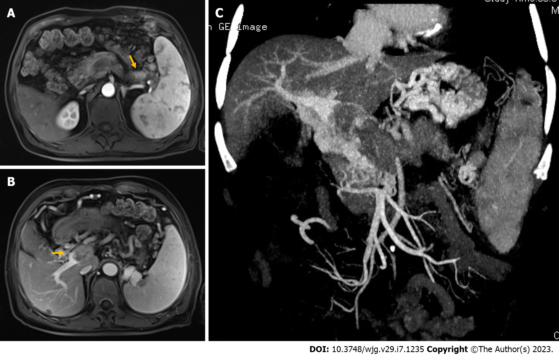
Figure 1 Representative magnetic resonance imaging and computed tomography image on admission.
A: Contrast-enhanced magnetic resonance imaging (MRI) axial view showing a well-enhanced tumor in the pancreatic tail; B: Axial view of contrast-enhanced MRI shows an intravascular filling defect (arrows) occupying the entire portal vein lumen; C: Computed tomography portal venography image showing extensive collateral venous circulation due to portal vein occlusion.
- Citation: Wang GC, Huang GJ, Zhang CQ, Ding Q. Percutaneous transhepatic intraportal biopsy using gastroscope biopsy forceps for diagnosis of a pancreatic neuroendocrine neoplasm: A case report. World J Gastroenterol 2023; 29(7): 1235-1242
- URL: https://www.wjgnet.com/1007-9327/full/v29/i7/1235.htm
- DOI: https://dx.doi.org/10.3748/wjg.v29.i7.1235


