Copyright
©The Author(s) 2023.
World J Gastroenterol. Feb 7, 2023; 29(5): 834-850
Published online Feb 7, 2023. doi: 10.3748/wjg.v29.i5.834
Published online Feb 7, 2023. doi: 10.3748/wjg.v29.i5.834
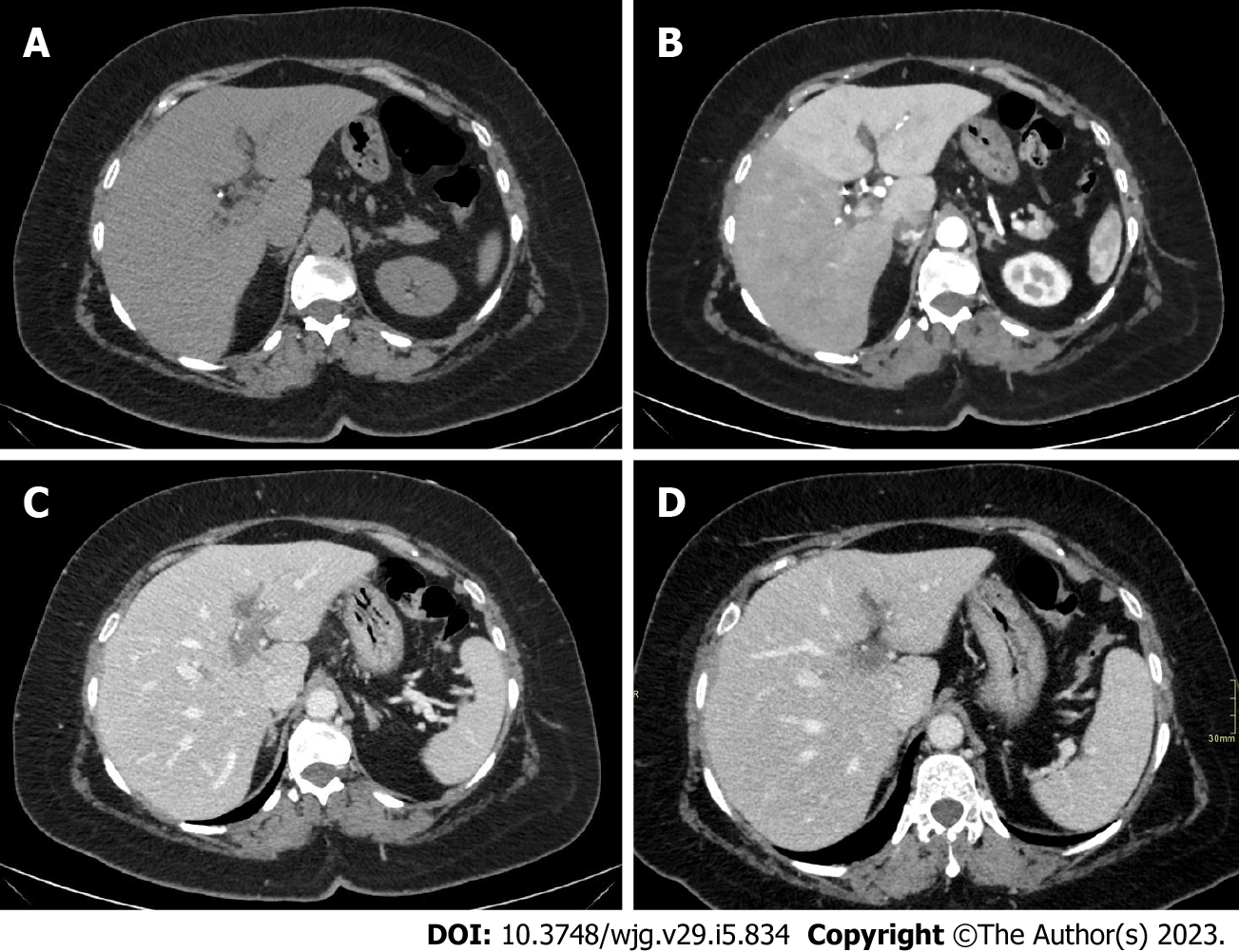
Figure 4 A 40-year-old men with coronavirus disease 2019 infection and marked respiratory symptoms, underwent contrast-enhanced computed tomography due to elevated liver and biliary enzymes.
A and B: On unenhanced phase (A) the liver is within normal limits. After the injection of contrast agent, on arterial phase (B) liver parenchyma shows inhomogeneous enhancement, with hypervascularization of the left liver lobe, as in case of transient hepatic attenuation differences; C: On the portal venous phase, the arterial hypervascularization fades to homogeneous enhancement and diffuse thrombosis of the left branch of the portal vein is demonstrated; D: After 6 mo the patient underwent contrast-enhanced computed tomography that demonstrated persistent portal vein thrombosis without venous collaterals.
- Citation: Ippolito D, Maino C, Vernuccio F, Cannella R, Inchingolo R, Dezio M, Faletti R, Bonaffini PA, Gatti M, Sironi S. Liver involvement in patients with COVID-19 infection: A comprehensive overview of diagnostic imaging features. World J Gastroenterol 2023; 29(5): 834-850
- URL: https://www.wjgnet.com/1007-9327/full/v29/i5/834.htm
- DOI: https://dx.doi.org/10.3748/wjg.v29.i5.834


