Copyright
©The Author(s) 2023.
World J Gastroenterol. Nov 21, 2023; 29(43): 5834-5847
Published online Nov 21, 2023. doi: 10.3748/wjg.v29.i43.5834
Published online Nov 21, 2023. doi: 10.3748/wjg.v29.i43.5834
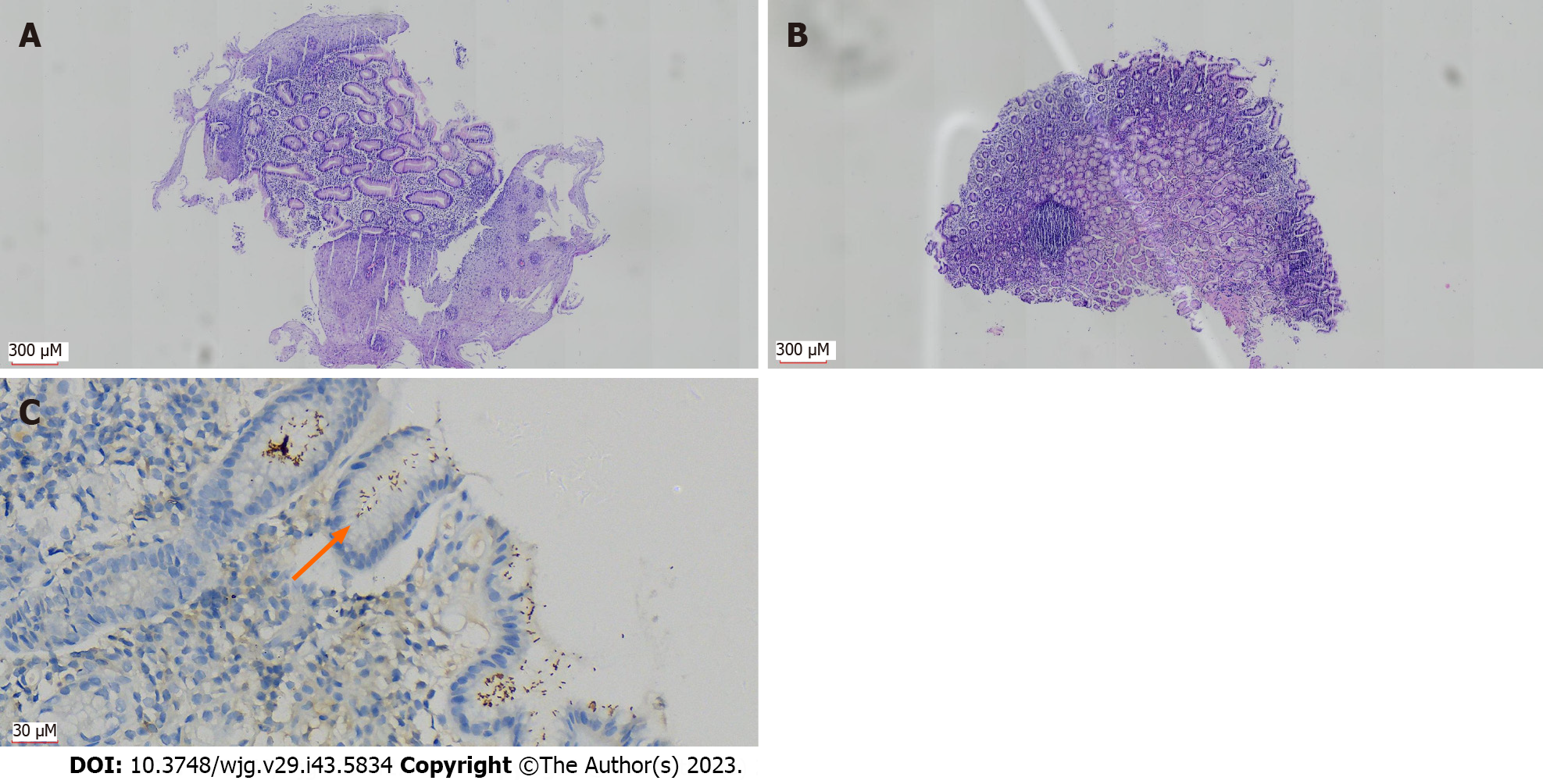
Figure 3 Patients with Barrett’s esophagus concurrent with Helicobacter pylori infection.
A: Changes with Barrett’s esophagus: Many hyperplastic glands in the esophageal wall, partially replacing the squamous epithelial mucosa [hematoxylin and eosin (H&E, 40 ×)]; B: Mild inflammation (H&E, 40 ×) in the gastric wall tissue biopsied simultaneously; C: Immunohistochemistry showing Helicobacter pylori (H. pylori) infection (DAB, 400 ×); orange arrow in C indicates distribution of H. pylori.
- Citation: Peng YH, Feng X, Zhou Z, Yang L, Shi YF. Helicobacter pylori infection in Xinjiang Uyghur Autonomous Region: Prevalence and analysis of related factors. World J Gastroenterol 2023; 29(43): 5834-5847
- URL: https://www.wjgnet.com/1007-9327/full/v29/i43/5834.htm
- DOI: https://dx.doi.org/10.3748/wjg.v29.i43.5834


