Copyright
©The Author(s) 2023.
World J Gastroenterol. Nov 21, 2023; 29(43): 5834-5847
Published online Nov 21, 2023. doi: 10.3748/wjg.v29.i43.5834
Published online Nov 21, 2023. doi: 10.3748/wjg.v29.i43.5834
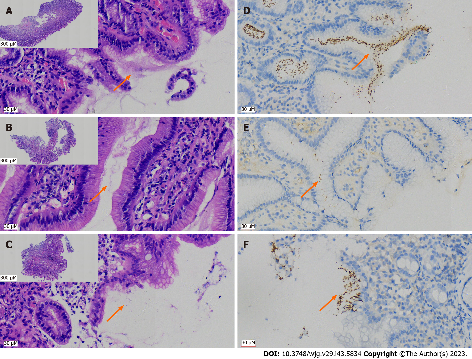
Figure 1 Immunohistochemical staining using antibodies against Helicobacter pylori has significant advantages over hematoxylin and eosin staining in the pathological tissue for gastroscopy biopsy.
A: Distribution of curved rod-shaped bacteria [hematoxylin and eosin (H&E, 400 ×)], on the surface of glands and in the mucus of typical Helicobacter pylori (H. pylori)-positive patients in H&E section; B: A case with low bacterial count of H. pylori (H&E, 400 ×); C: A case with atypical H. pylori morphology (mainly coccoid rather than rod-shaped), which was difficult to identify (H&E, 400 ×), lower magnification figures of A-C are in the upper left corner (H&E, 40 ×); D: Immunohistochemistry (IHC) with antibodies against H. pylori indicated a large number of bacteria, exhibiting brown yellow deep staining (DAB, high magnification, one case the same as in A); E: IHC displayed H. pylori bacilli more clearly than H&E due to a lower bacterial count (DAB staining, high magnification, one case the same as in B); F: Atypical coccoid H. pylori were clearly present and confirmed by IHC (DAB staining, high magnification, one case the same as in C); orange arrows in A-F indicate distribution of H. pylori.
- Citation: Peng YH, Feng X, Zhou Z, Yang L, Shi YF. Helicobacter pylori infection in Xinjiang Uyghur Autonomous Region: Prevalence and analysis of related factors. World J Gastroenterol 2023; 29(43): 5834-5847
- URL: https://www.wjgnet.com/1007-9327/full/v29/i43/5834.htm
- DOI: https://dx.doi.org/10.3748/wjg.v29.i43.5834


