Copyright
©The Author(s) 2023.
World J Gastroenterol. Oct 28, 2023; 29(40): 5543-5556
Published online Oct 28, 2023. doi: 10.3748/wjg.v29.i40.5543
Published online Oct 28, 2023. doi: 10.3748/wjg.v29.i40.5543
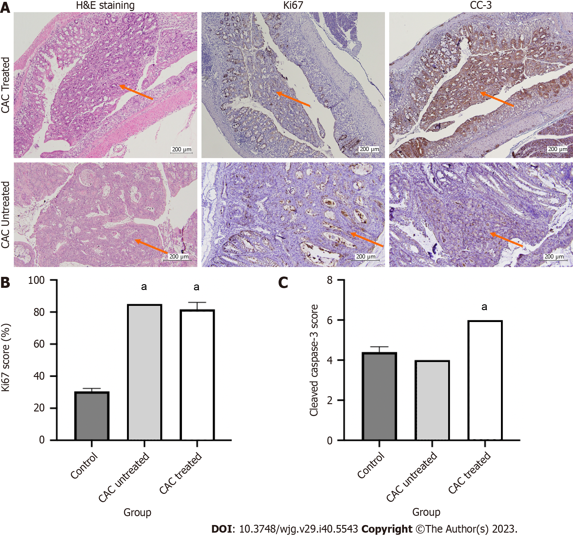
Figure 7 Immunohistochemistry analysis on Ki67 and cleaved caspase-3 markers on colitis-associated cancer-mice model.
A: H&E staining, Ki67 and cleaved caspase-3 (CC-3) staining; B and C: Ki67 and CC-3 staining exhibited lower proliferative and higher apoptotic cells on the buparlisib-treated colitis-associated cancer (CAC) mice model compared to the untreated CAC mice. aP < 0.05 (compared to the control group); data are presented as mean ± SEM; one-way ANOVA was employed; n = 4-8. The red arrow showed the tumor area. Magnification is 100 ×. CAC: Colitis-associated cancer; CC-3: Cleaved caspase-3.
- Citation: Razali NN, Raja Ali RA, Muhammad Nawawi KN, Yahaya A, Mohd Rathi ND, Mokhtar NM. Roles of phosphatidylinositol-3-kinases signaling pathway in inflammation-related cancer: Impact of rs10889677 variant and buparlisib in colitis-associated cancer. World J Gastroenterol 2023; 29(40): 5543-5556
- URL: https://www.wjgnet.com/1007-9327/full/v29/i40/5543.htm
- DOI: https://dx.doi.org/10.3748/wjg.v29.i40.5543


