Copyright
©The Author(s) 2023.
World J Gastroenterol. Aug 28, 2023; 29(32): 4860-4872
Published online Aug 28, 2023. doi: 10.3748/wjg.v29.i32.4860
Published online Aug 28, 2023. doi: 10.3748/wjg.v29.i32.4860
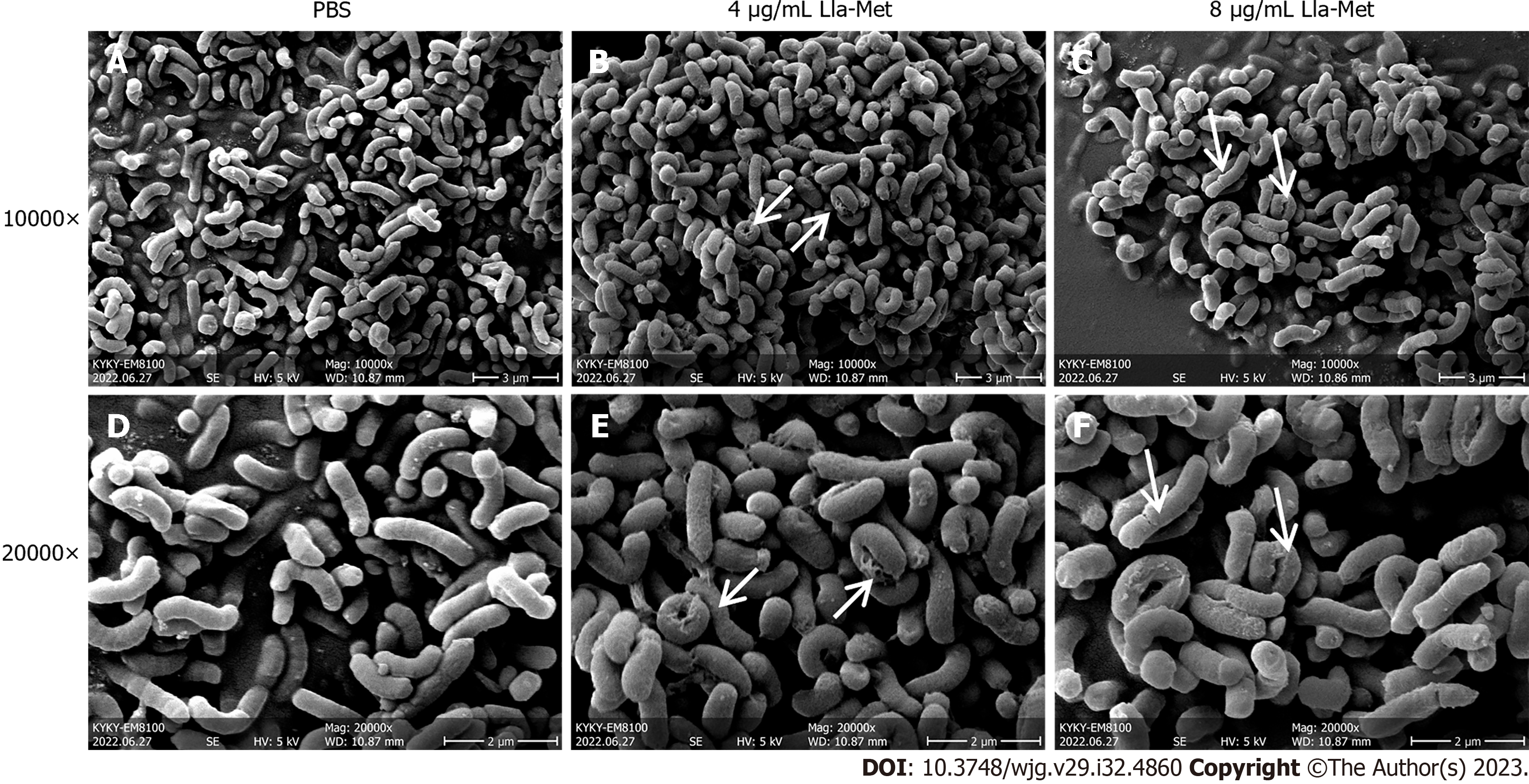
Figure 1 The effect of linolenic acid-metronidazole on Helicobacter pylori morphology.
A: The control group shows the cell morphology at × 10000 magnification; B: The appearance of Helicobacter pylori (H. pylori) in linolenic acid-metronidazole concentrations of 4 μg/mL shows the cell morphology at × 10000 magnification; C: The appearance of H. pylori in linolenic acid-metronidazole concentrations of 8 μg/mL shows the cell morphology at × 10000 magnification; D: The control group shows the cell morphology at × 20000 magnification; E: The appearance of H. pylori in linolenic acid-metronidazole concentrations of 4 μg/mL shows the cell morphology at × 20000 magnification; F: The appearance of H. pylori in linolenic acid-metronidazole concentrations of 8 μg/mL shows the cell morphology at × 20000 magnification. The arrow points to the cell damage. Roughness, swelling, breakages on the cell surface, etc., are shown. PBS: Phosphate buffered saline; Lla-Met: Linolenic acid-metronidazole.
- Citation: Zhou WT, Dai YY, Liao LJ, Yang SX, Chen H, Huang L, Zhao JL, Huang YQ. Linolenic acid-metronidazole inhibits the growth of Helicobacter pylori through oxidation. World J Gastroenterol 2023; 29(32): 4860-4872
- URL: https://www.wjgnet.com/1007-9327/full/v29/i32/4860.htm
- DOI: https://dx.doi.org/10.3748/wjg.v29.i32.4860


