Copyright
©The Author(s) 2023.
World J Gastroenterol. Jul 21, 2023; 29(27): 4222-4235
Published online Jul 21, 2023. doi: 10.3748/wjg.v29.i27.4222
Published online Jul 21, 2023. doi: 10.3748/wjg.v29.i27.4222
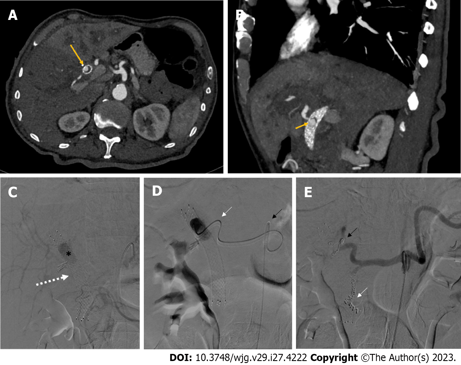
Figure 7 Non-variceal upper gastrointestinal bleeding due to hemobilia.
A and B: Contrast-enhanced computed tomography axial scan in the arterial phase (A) and its maximum intensity projection oblique-sagittal reconstruction (B) showing a pseudoaneurysm in the lumen of the metallic biliary stent (long arrow, A), arising from the hepatic artery (short arrow, B); C: Delayed phase celiac angiography showing a pseudoaneurysm (black asterisk) into the lumen of a metallic biliary stent (white arrow); D: The pseudoaneurysm of the proper hepatic artery was reached with a 2.7 Fr microcatheter (white arrow) coaxially through a 5 Fr catheter (black arrow) positioned into the celiac trunk; E: Successful coil embolization of both the proper hepatic artery (black arrow) and the gastroduodenal artery (white arrow), in order to avoid recanalization.
- Citation: Martino A, Di Serafino M, Orsini L, Giurazza F, Fiorentino R, Crolla E, Campione S, Molino C, Romano L, Lombardi G. Rare causes of acute non-variceal upper gastrointestinal bleeding: A comprehensive review. World J Gastroenterol 2023; 29(27): 4222-4235
- URL: https://www.wjgnet.com/1007-9327/full/v29/i27/4222.htm
- DOI: https://dx.doi.org/10.3748/wjg.v29.i27.4222


