Copyright
©The Author(s) 2023.
World J Gastroenterol. Jul 21, 2023; 29(27): 4222-4235
Published online Jul 21, 2023. doi: 10.3748/wjg.v29.i27.4222
Published online Jul 21, 2023. doi: 10.3748/wjg.v29.i27.4222
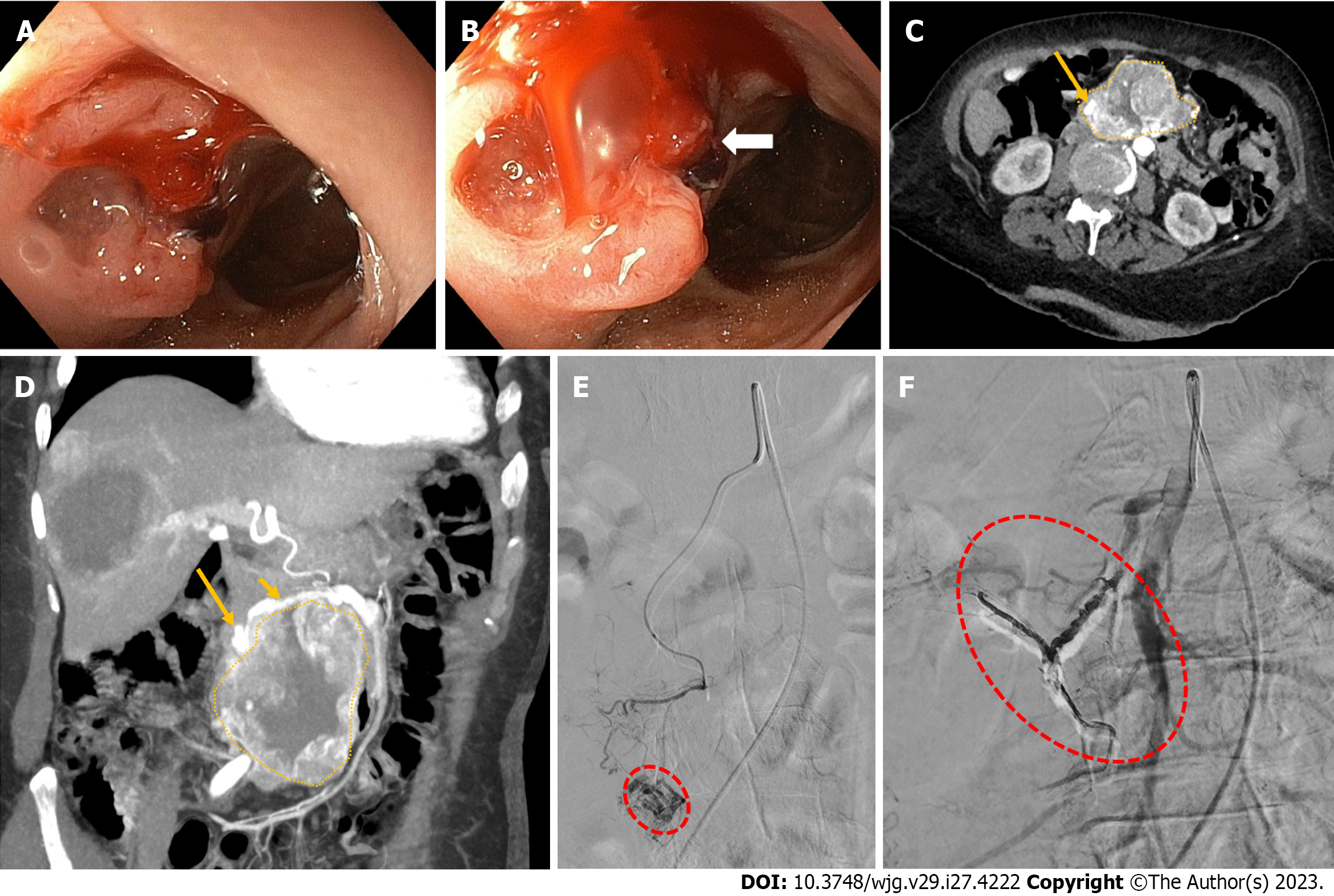
Figure 2 Non-variceal upper gastrointestinal bleeding due rupture of a superior mesenteric artery pseudoaneurysm into the duodenum.
A and B: Upper endoscopy showing a bulging, focally ulcerated, lesion in the second-third part of the duodenum, with a suspected small fistulous orifice on its surface (arrow); C and D: Contrast-enhanced computed tomography axial scan in the arterial phase (C) and its maximum intensity projection oblique-coronal reconstruction (D) showing a large lesion (dotted area, C and D) arising from the second-third part of the duodenum, with peripheral vascularity and a pseudoaneurysm (long arrow, C and D) arising from branches of the superior mesenteric artery (short arrow, B); E: Superselective superior mesenteric artery angiography showing a pseudoaneurysm (red dotted circle) fed by the inferior pancreaticoduodenal artery; F: Successful endovascular embolization by the use of cohesive glue (Onyx-18®, Medtronic, Dublin, Ireland) with disappearance of the pseudoaneurysm and visibility of the Onyx-18® cast (red dotted circle).
- Citation: Martino A, Di Serafino M, Orsini L, Giurazza F, Fiorentino R, Crolla E, Campione S, Molino C, Romano L, Lombardi G. Rare causes of acute non-variceal upper gastrointestinal bleeding: A comprehensive review. World J Gastroenterol 2023; 29(27): 4222-4235
- URL: https://www.wjgnet.com/1007-9327/full/v29/i27/4222.htm
- DOI: https://dx.doi.org/10.3748/wjg.v29.i27.4222


