Copyright
©The Author(s) 2023.
World J Gastroenterol. Jun 14, 2023; 29(22): 3440-3468
Published online Jun 14, 2023. doi: 10.3748/wjg.v29.i22.3440
Published online Jun 14, 2023. doi: 10.3748/wjg.v29.i22.3440
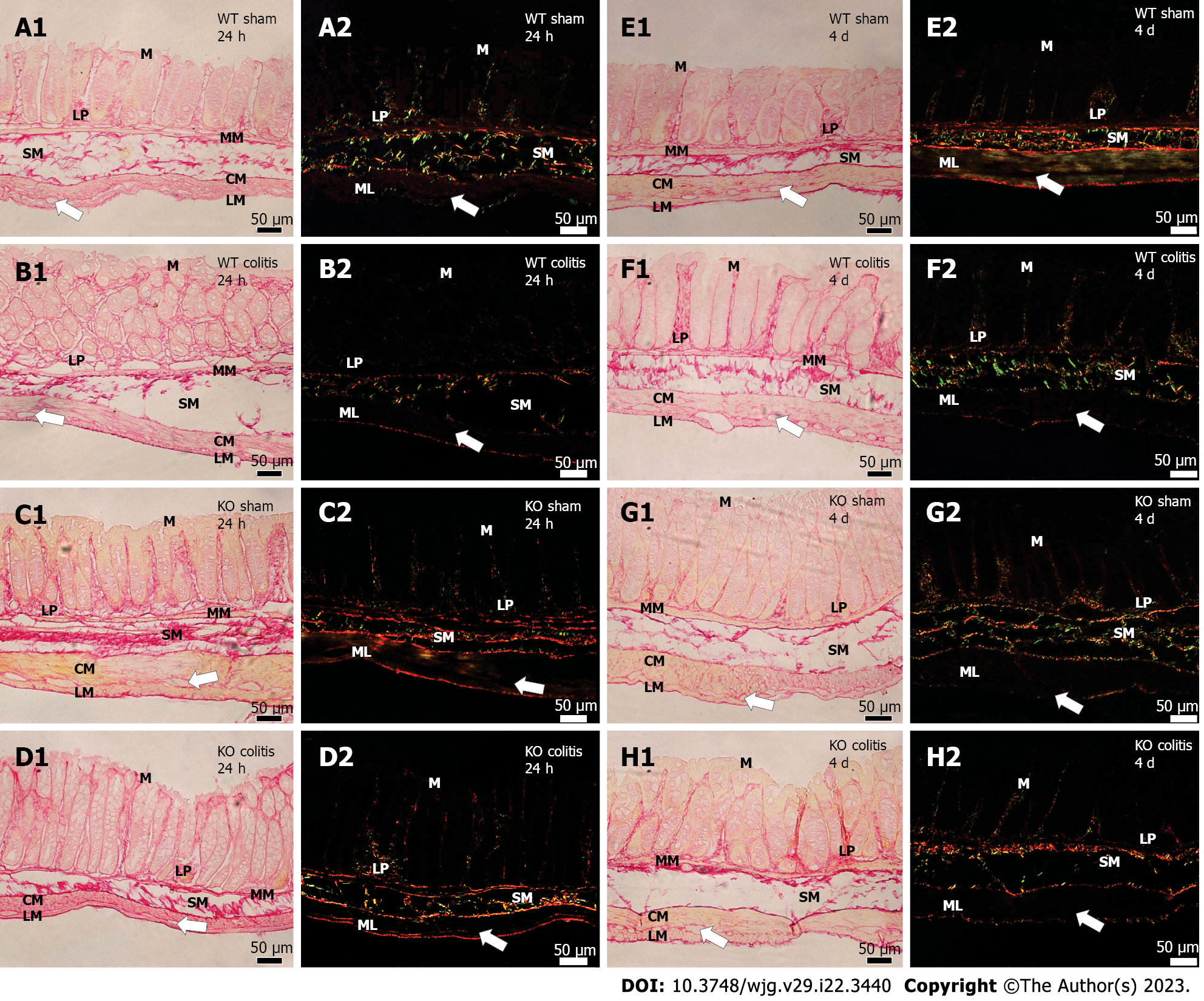
Figure 13 Photomicrograph of picrosirius red-stained transverse sections of the distal colons of mice of different groups.
A1 and A2: Wild-type (WT) sham 24 h; B1 and B2: WT colitis 24 h; C1 and C2: Knockout (KO) sham 24 h; D1 and D2: KO colitis 24 h; E1 and E2: WT sham 4 d; F1 and F2: WT colitis 4 d; G1 and G2: KO sham 4 d; H1 and H2: KO colitis 4 d groups taken under an optical light (red) or polarized light (green) microscope. Thick collagen fibers (red) in the lamina propria and submucosal layer, intermingled with and involving the muscle layer, and circumscribing the myenteric ganglia were observed in the sham groups. In the WT colitis group, there was a decrease in the number of these fibers in the submucosal layer and around the myenteric ganglia, an increase in the number and disorganization of these fibers in the lamina propria and accumulation of these fibers in the subepithelial region. Tissue was better preserved in the KO colitis group. M: Mucosal layer; LP: Lamina propria; MM: Mucosal muscle; SM: Submucosal layer; ML: Muscle layer; CM: Circular musculature; LM: Longitudinal musculature; WT: Wild-type; KO: Knockout. White arrows mean myenteric ganglion.
- Citation: Magalhães HIR, Machado FA, Souza RF, Caetano MAF, Figliuolo VR, Coutinho-Silva R, Castelucci P. Study of the roles of caspase-3 and nuclear factor kappa B in myenteric neurons in a P2X7 receptor knockout mouse model of ulcerative colitis. World J Gastroenterol 2023; 29(22): 3440-3468
- URL: https://www.wjgnet.com/1007-9327/full/v29/i22/3440.htm
- DOI: https://dx.doi.org/10.3748/wjg.v29.i22.3440


