Copyright
©The Author(s) 2023.
World J Gastroenterol. Jun 7, 2023; 29(21): 3257-3268
Published online Jun 7, 2023. doi: 10.3748/wjg.v29.i21.3257
Published online Jun 7, 2023. doi: 10.3748/wjg.v29.i21.3257
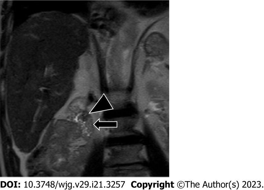
Figure 9 Enlarged ampullary papilla occurring years after liver transplantation and causing minimal cholestasis.
T2-weighted imaging in the coronal plane demonstrates an enlarged ampullary papilla (arrowhead) protruding in the duodenal lumen (arrow). Ultrasonography-endoscopy confirmed the enlarged ampullary papilla and biopsy was performed, which excluded malignancy and confirmed the diagnosis of papillary stenosis (i.e. sphincter of Oddi dysfunction); sphincterotomy was then performed.
- Citation: Vernuccio F, Mercante I, Tong XX, Crimì F, Cillo U, Quaia E. Biliary complications after liver transplantation: A computed tomography and magnetic resonance imaging pictorial review. World J Gastroenterol 2023; 29(21): 3257-3268
- URL: https://www.wjgnet.com/1007-9327/full/v29/i21/3257.htm
- DOI: https://dx.doi.org/10.3748/wjg.v29.i21.3257


