Copyright
©The Author(s) 2023.
World J Gastroenterol. Mar 28, 2023; 29(12): 1852-1862
Published online Mar 28, 2023. doi: 10.3748/wjg.v29.i12.1852
Published online Mar 28, 2023. doi: 10.3748/wjg.v29.i12.1852
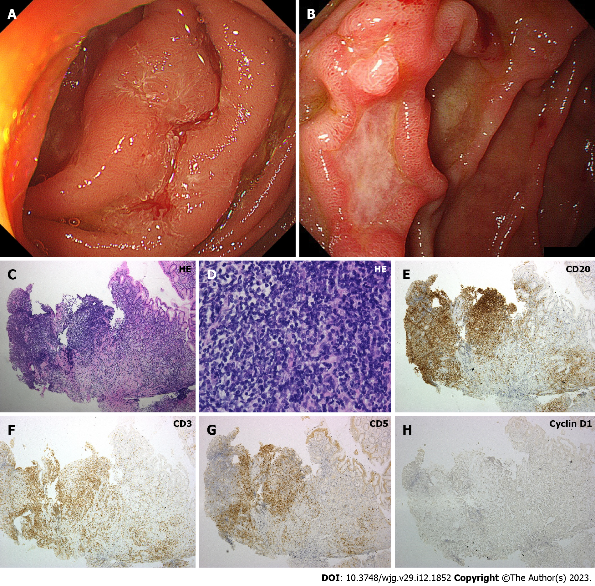
Figure 5 Representative endoscopic and pathological images of duodenal MALT lymphoma (Cases 8 and 9).
A: Case 8. A protruded lesion mimicking hypertrophic folds is observed in the duodenum; B: Case 9. Esophagogastroduodenoscopy shows ulcerative tumor in the duodenum; C–H: Pathological images of Case 9. On hematoxylin and eosin stain, small to medium-sized lymphoma cells are predominant (C: × 10, D: × 40). Neoplastic cells are positive for CD20, while negative for CD3, CD5, and Cyclin D1. HE: Hematoxylin and eosin.
- Citation: Iwamuro M, Tanaka T, Okada H. Review of lymphoma in the duodenum: An update of diagnosis and management. World J Gastroenterol 2023; 29(12): 1852-1862
- URL: https://www.wjgnet.com/1007-9327/full/v29/i12/1852.htm
- DOI: https://dx.doi.org/10.3748/wjg.v29.i12.1852


