Copyright
©The Author(s) 2022.
World J Gastroenterol. Feb 28, 2022; 28(8): 868-877
Published online Feb 28, 2022. doi: 10.3748/wjg.v28.i8.868
Published online Feb 28, 2022. doi: 10.3748/wjg.v28.i8.868
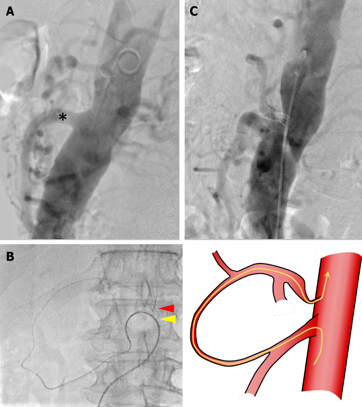
Figure 2 Preoperative endovascular stenting.
A: In preoperative aortography, the superior mesenteric artery (SMA) was visualized immediately, but the celiac axis (CA) was not visualized. Black asterisks: SMA; B: The microguidewire reached the CA via a collateral pathway from the SMA using a triple coaxial system; C: Final aortography confirmed CA patency and antegrade blood flow. Red arrowhead: Root of the CA; yellow arrowhead: Root of the SMA; yellow line: Running of wire.
- Citation: Yoshida E, Kimura Y, Kyuno T, Kawagishi R, Sato K, Kono T, Chiba T, Kimura T, Yonezawa H, Funato O, Kobayashi M, Murakami K, Takagane A, Takemasa I. Treatment strategy for pancreatic head cancer with celiac axis stenosis in pancreaticoduodenectomy: A case report and review of literature. World J Gastroenterol 2022; 28(8): 868-877
- URL: https://www.wjgnet.com/1007-9327/full/v28/i8/868.htm
- DOI: https://dx.doi.org/10.3748/wjg.v28.i8.868


