Copyright
©The Author(s) 2022.
World J Gastroenterol. Feb 7, 2022; 28(5): 594-601
Published online Feb 7, 2022. doi: 10.3748/wjg.v28.i5.594
Published online Feb 7, 2022. doi: 10.3748/wjg.v28.i5.594
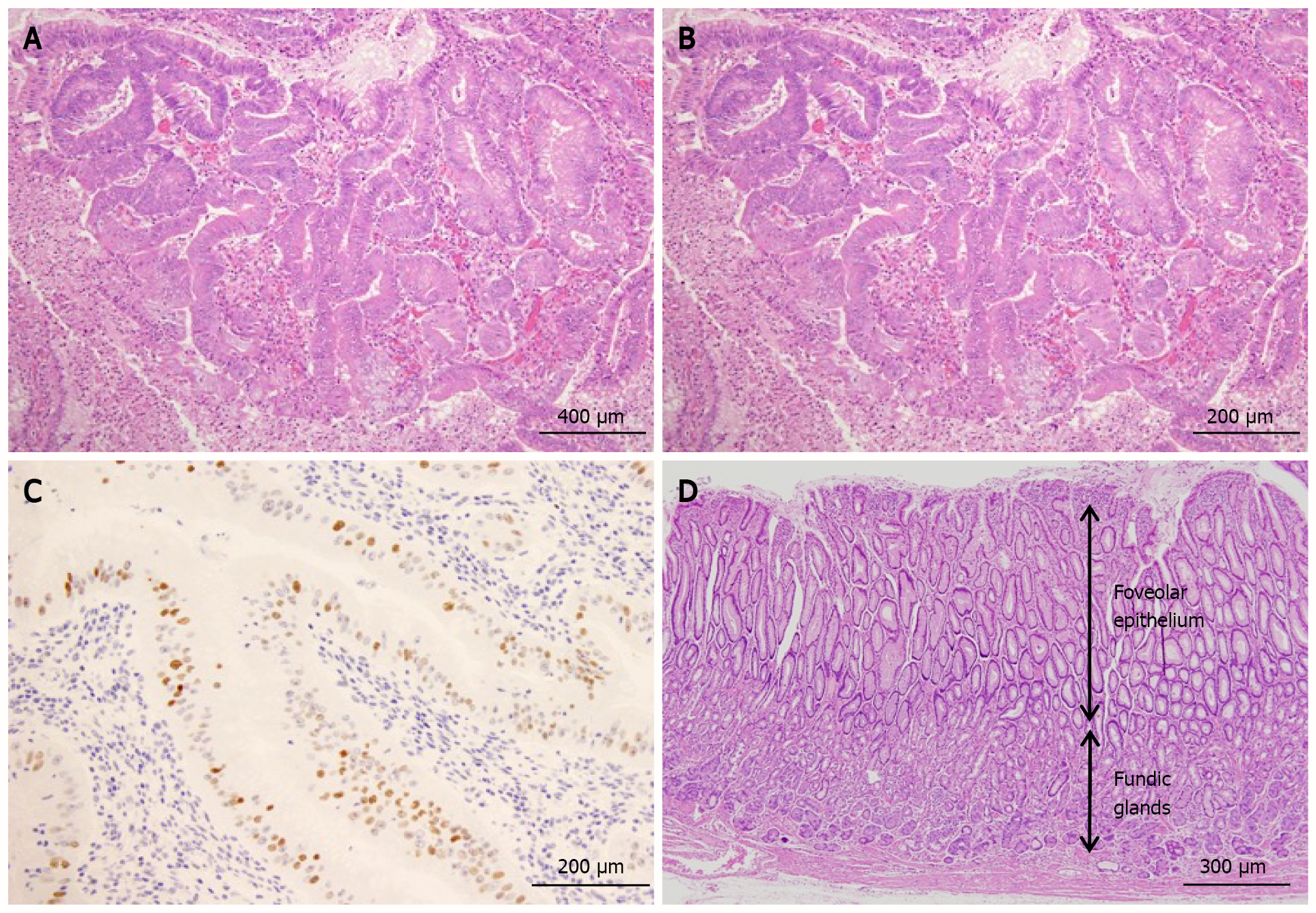
Figure 4 Histological findings.
A: The protruding lesion exhibits proliferation of atypical glands with irregular tubular structures (HE; × 20); B: High magnification shows proliferation of atypical cells and enlarged nuclei (irregular or oval-shaped) in the irregular tubular structure (HE; × 20); C: Scattered positive image (in nuclei) in the region (p53; × 20); D: Foveolar hyperplasia and atrophy of the proper gastric glands in non-cancerous area, consistent with Ménétrier’s disease (HE; × 20).
- Citation: Fukushi K, Goda K, Kino H, Kondo M, Kanazawa M, Kashima K, Kanamori A, Abe K, Suzuki T, Tominaga K, Yamagishi H, Irisawa A. Curative resection with endoscopic submucosal dissection of early gastric cancer in Helicobacter pylori-negative Ménétrier’s disease: A case report. World J Gastroenterol 2022; 28(5): 594-601
- URL: https://www.wjgnet.com/1007-9327/full/v28/i5/594.htm
- DOI: https://dx.doi.org/10.3748/wjg.v28.i5.594


