Copyright
©The Author(s) 2022.
World J Gastroenterol. Feb 7, 2022; 28(5): 594-601
Published online Feb 7, 2022. doi: 10.3748/wjg.v28.i5.594
Published online Feb 7, 2022. doi: 10.3748/wjg.v28.i5.594
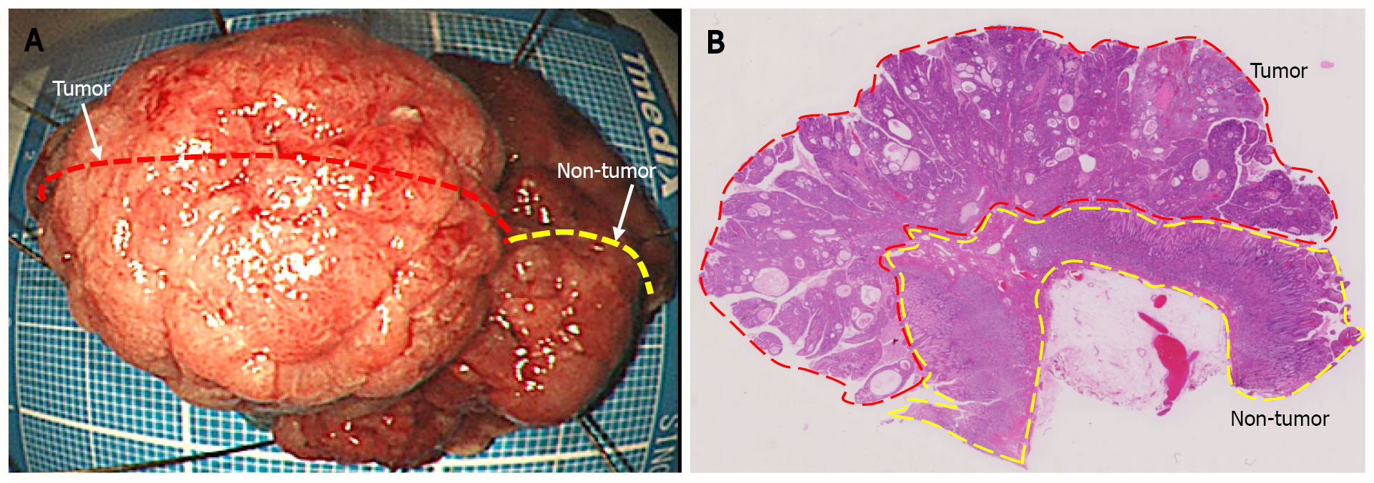
Figure 3 Endoscopic submucosal dissection specimen.
A: 45 mm × 38 mm resection specimen comprising a broad-based protruding lesion with nodular surface and uninvolved (non-tumor) tissue with giant rugae; B: Loupe image shows that the well-differentiated tubular adenocarcinoma was confined to the mucosal layer, with no lymphovascular invasion (ly0/v0) or ulceration (UL0), and both the lateral and vertical margins were negative.
- Citation: Fukushi K, Goda K, Kino H, Kondo M, Kanazawa M, Kashima K, Kanamori A, Abe K, Suzuki T, Tominaga K, Yamagishi H, Irisawa A. Curative resection with endoscopic submucosal dissection of early gastric cancer in Helicobacter pylori-negative Ménétrier’s disease: A case report. World J Gastroenterol 2022; 28(5): 594-601
- URL: https://www.wjgnet.com/1007-9327/full/v28/i5/594.htm
- DOI: https://dx.doi.org/10.3748/wjg.v28.i5.594


