Copyright
©The Author(s) 2022.
World J Gastroenterol. Dec 28, 2022; 28(48): 6935-6949
Published online Dec 28, 2022. doi: 10.3748/wjg.v28.i48.6935
Published online Dec 28, 2022. doi: 10.3748/wjg.v28.i48.6935
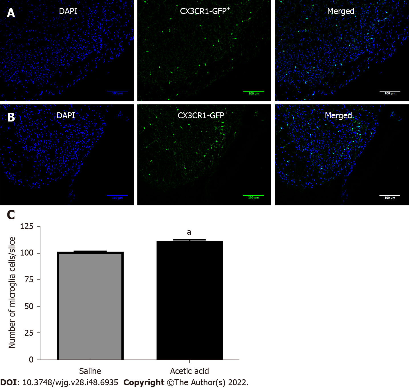
Figure 4 Number of microglia cells in the spinal cord.
A and B: Microglial cells in the dorsal horn of the L6-S1 level of the spinal cord were stained green [green fluorescent protein on the fractalkine receptor-positive (CX3CR1gfp+) mice], while nuclei appear in blue (DAPI) at day 7 after intravesical injections of saline (NaCl 0.9%) (A) or acetic acid 0.75% (B); C: The number of microglial cells per field was compared between the two groups using a Mann-Whitney bilateral test (n = 5 per group, average of 23 slices analyzed per mouse). aP < 0.05. Results are expressed as the mean ± standard error of the mean.
- Citation: Atmani K, Wuestenberghs F, Baron M, Bouleté I, Guérin C, Bahlouli W, Vaudry D, do Rego JC, Cornu JN, Leroi AM, Coëffier M, Meleine M, Gourcerol G. Bladder-colon chronic cross-sensitization involves neuro-glial pathways in male mice. World J Gastroenterol 2022; 28(48): 6935-6949
- URL: https://www.wjgnet.com/1007-9327/full/v28/i48/6935.htm
- DOI: https://dx.doi.org/10.3748/wjg.v28.i48.6935


