Copyright
©The Author(s) 2022.
World J Gastroenterol. Nov 28, 2022; 28(44): 6206-6212
Published online Nov 28, 2022. doi: 10.3748/wjg.v28.i44.6206
Published online Nov 28, 2022. doi: 10.3748/wjg.v28.i44.6206
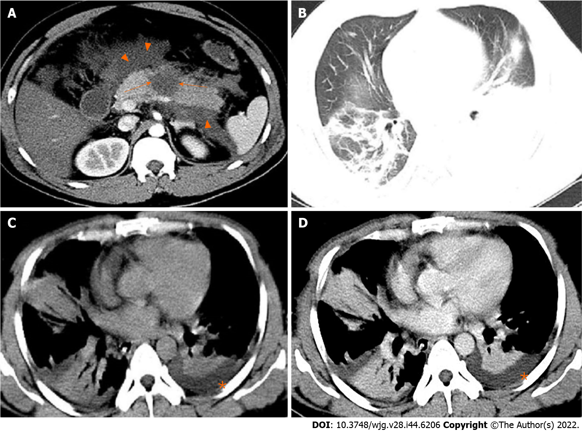
Figure 1 A 31-year-old man with severe acute pancreatitis complicated with acute respiratory distress syndrome.
A: Axial contrast-enhanced computed tomography image in the arterial phase shows flake parenchymal necrosis (arrows) in the region of body of the pancreas, as well as extensive heterogeneous collections (acute necrotic collections) in the peripancreatic and the left pararenal anterior spaces (arrowheads); B: Lung window; C: Mediastinal window; D: Chest axial contrast-enhanced venous phase image. The three images show partial pulmonary consolidation in the middle and lower lobes of the right lung, in which bronchial inflation signs can be seen, and partial consolidation with partial atelectasis in the lower lobe of the left lung caused by external pressure of pleural effusion (asterisks).
- Citation: Song LJ, Xiao B. Medical imaging for pancreatic diseases: Prediction of severe acute pancreatitis complicated with acute respiratory distress syndrome. World J Gastroenterol 2022; 28(44): 6206-6212
- URL: https://www.wjgnet.com/1007-9327/full/v28/i44/6206.htm
- DOI: https://dx.doi.org/10.3748/wjg.v28.i44.6206


