Copyright
©The Author(s) 2022.
World J Gastroenterol. Oct 28, 2022; 28(40): 5827-5844
Published online Oct 28, 2022. doi: 10.3748/wjg.v28.i40.5827
Published online Oct 28, 2022. doi: 10.3748/wjg.v28.i40.5827
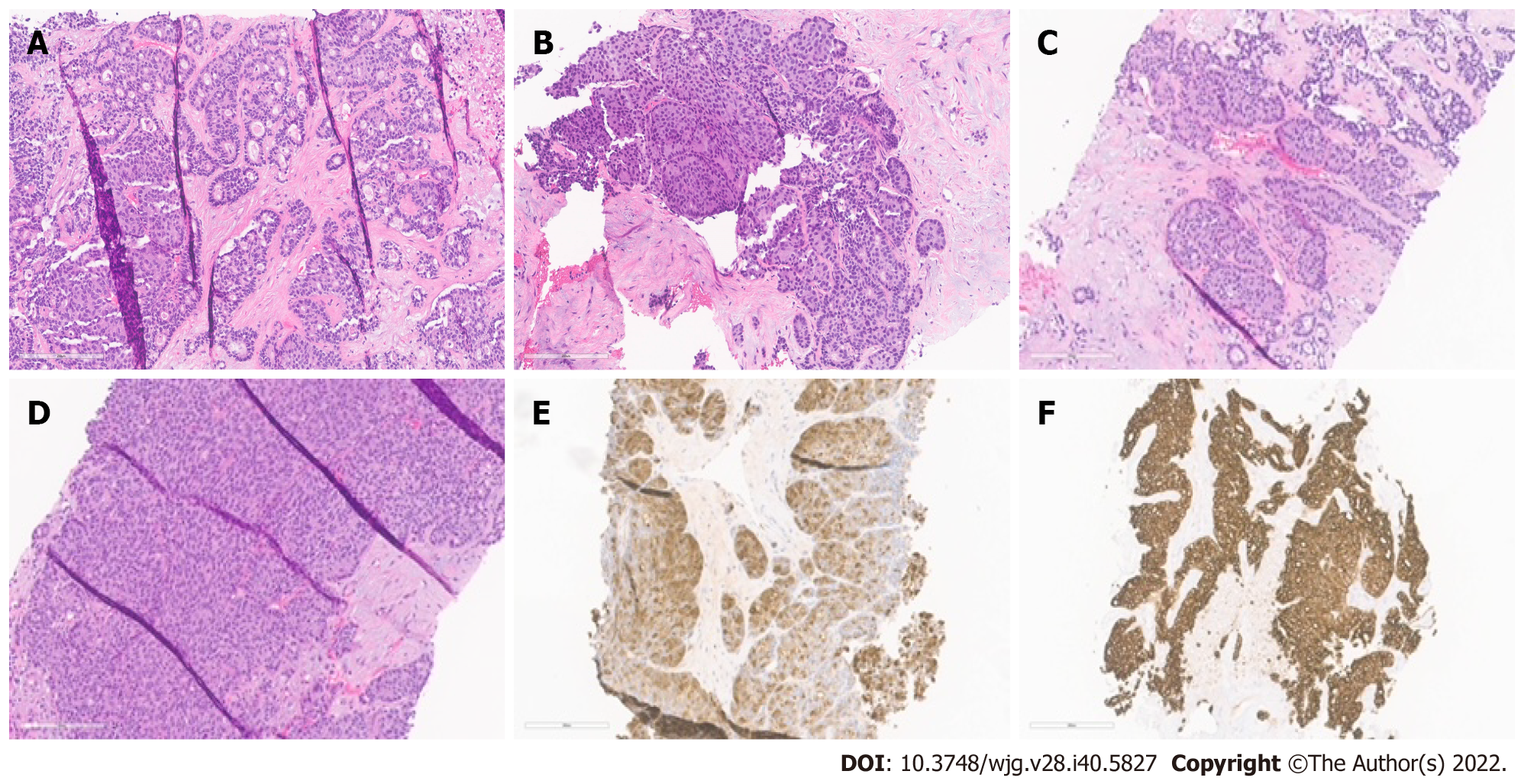
Figure 1 Histopathological patterns.
A: Acinar cell carcinoma with the predominant acinar pattern. The acinar pattern is characterized by structures resembling normal acini, with small lumina and cells arranged in a monolayer with basally located nuclei; B: Acinar cell carcinoma with the predominant glandular pattern. Acinar structures with dilated lumina characterize the glandular pattern; C: Acinar cell carcinoma with the predominant trabecular pattern. The trabecular pattern is characterized by ribbons of cells resembling those of pancreatic neuroendocrine tumors; D: Acinar cell carcinoma with a predominant solid pattern. Large sheets of cells characterize the solid pattern without lumina; E: Acinar cell carcinoma, immunohistochemical staining for trypsin; F: Acinar cell carcinoma, immunohistochemical staining for cytokeratin 7.
- Citation: Calimano-Ramirez LF, Daoud T, Gopireddy DR, Morani AC, Waters R, Gumus K, Klekers AR, Bhosale PR, Virarkar MK. Pancreatic acinar cell carcinoma: A comprehensive review. World J Gastroenterol 2022; 28(40): 5827-5844
- URL: https://www.wjgnet.com/1007-9327/full/v28/i40/5827.htm
- DOI: https://dx.doi.org/10.3748/wjg.v28.i40.5827


