Copyright
©The Author(s) 2022.
World J Gastroenterol. Sep 28, 2022; 28(36): 5313-5323
Published online Sep 28, 2022. doi: 10.3748/wjg.v28.i36.5313
Published online Sep 28, 2022. doi: 10.3748/wjg.v28.i36.5313
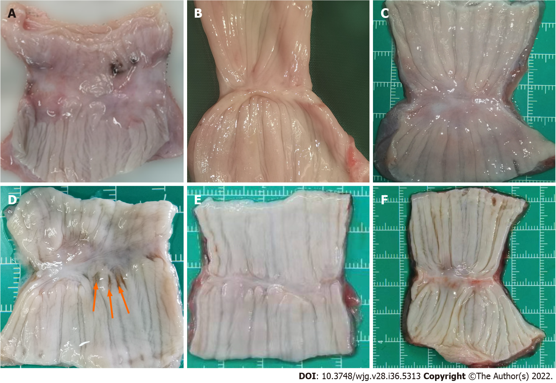
Figure 4 Gross appearance of the anastomosis in magnetic compression anastomosis and hand-sewn group.
A: The tissue of magnetic compression anastomosis (MCA) group is thin at 1 mo, but the mucous membrane is intact; B: Anastomotic tissue of the MCA group at 3 mo; C: At 6 mo, the tissue becomes smooth and flat in the MCA group. There is little difference from normal tissue; D: The tissue of the hand-sewn anastomotic stoma is incomplete, and the mucous membrane has a small pinhole at 1 mo, which was caused by the suture needle. Red arrow indicates; E: Anastomotic tissue of the hand-sewn group at 3 mo; F: Anastomotic tissue of the hand-sewn group at 6 mo.
- Citation: Xu XH, Lv Y, Liu SQ, Cui XH, Suo RY. Esophageal magnetic compression anastomosis in dogs. World J Gastroenterol 2022; 28(36): 5313-5323
- URL: https://www.wjgnet.com/1007-9327/full/v28/i36/5313.htm
- DOI: https://dx.doi.org/10.3748/wjg.v28.i36.5313


