Copyright
©The Author(s) 2022.
World J Gastroenterol. Sep 7, 2022; 28(33): 4812-4822
Published online Sep 7, 2022. doi: 10.3748/wjg.v28.i33.4812
Published online Sep 7, 2022. doi: 10.3748/wjg.v28.i33.4812
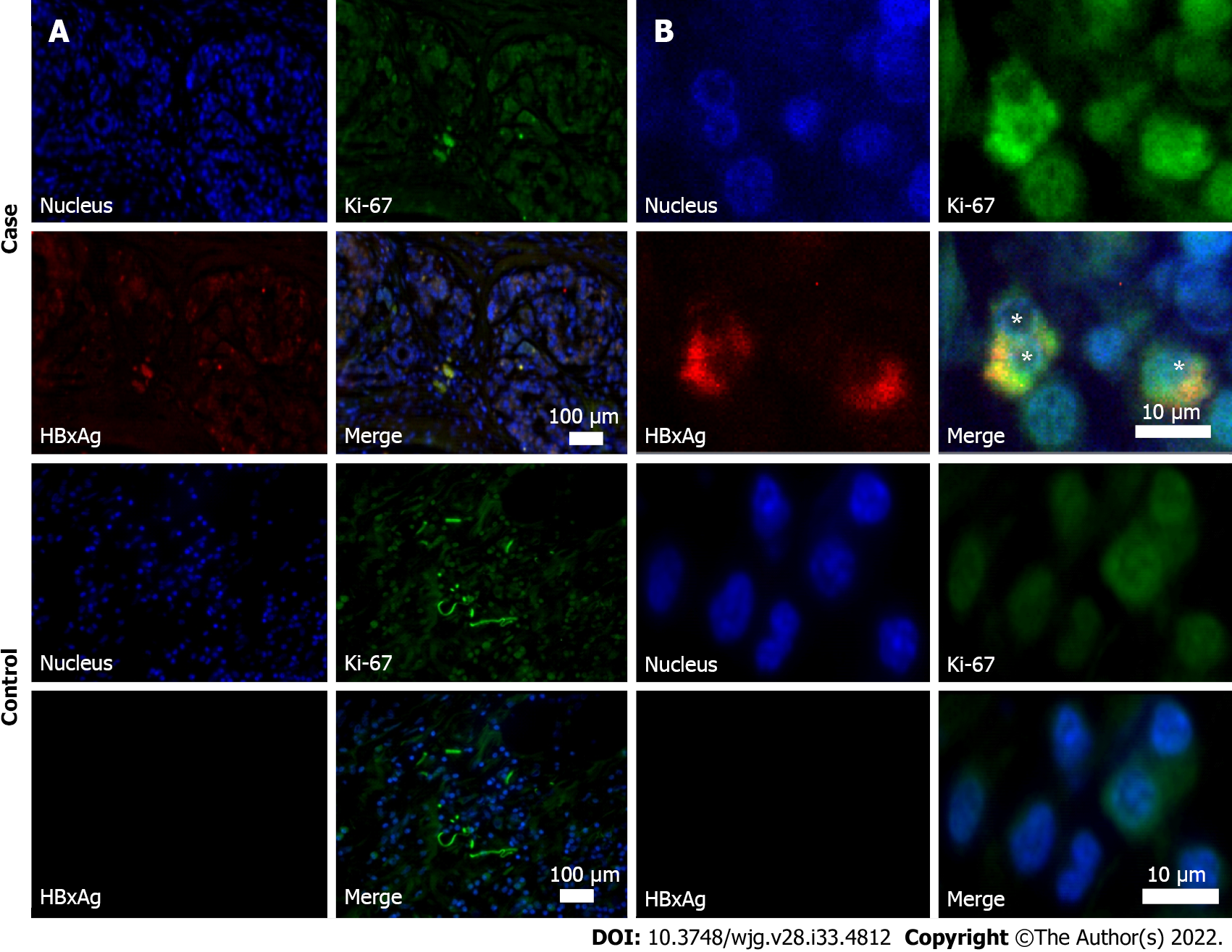
Figure 2 Immunohistochemistry of resected pancreatic tumor tissues (obtained during surgery).
Case: Anti-HBc-positive patient with pancreatic ductal adenocarcinoma. Control: Patient with pancreatic ductal adenocarcinoma, who was negative for hepatitis B virus biomarkers. A: Images at magnification × 10; B: Images at magnification × 100. Samples were stained for Ki-67 protein (green fluorescence) and X antigen of hepatitis B virus of (HBxAg) (red fluorescence). Cell nuclei were counterstained by Hoechst33342 dye (blue). Asterisks indicate HBxAg/Ki-67 co-stained cells.
- Citation: Batskikh S, Morozov S, Dorofeev A, Borunova Z, Kostyushev D, Brezgin S, Kostyusheva A, Chulanov V. Previous hepatitis B viral infection–an underestimated cause of pancreatic cancer. World J Gastroenterol 2022; 28(33): 4812-4822
- URL: https://www.wjgnet.com/1007-9327/full/v28/i33/4812.htm
- DOI: https://dx.doi.org/10.3748/wjg.v28.i33.4812


