Copyright
©The Author(s) 2022.
World J Gastroenterol. Aug 28, 2022; 28(32): 4540-4556
Published online Aug 28, 2022. doi: 10.3748/wjg.v28.i32.4540
Published online Aug 28, 2022. doi: 10.3748/wjg.v28.i32.4540
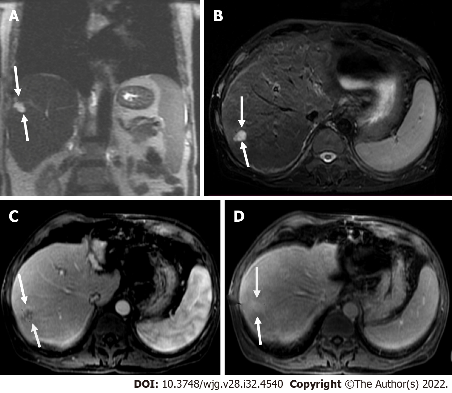
Figure 9 Coronal T2 weighted image and axial short tau inversion recovery image.
A and B: Coronal T2 weighted image (T2WI) (A) and axial short tau inversion recovery image (B) showed a 2 cm lesion in segment VII with high signal intensity (bright) within a nodular liver consistent with cirrhosis; C: Post-contrast image showed nodular peripheral enhancement; D: On delay image, the lesion showed progressive filling. In progressive cirrhosis, hemangiomas are likely to decrease in size, become more fibrotic, and are difficult to diagnose radiologically. However, the bright signal on T2WI and the enhancement pattern was pathognomonic for hemangioma of the liver consistent with LR-2.
- Citation: Liava C, Sinakos E, Papadopoulou E, Giannakopoulou L, Potsi S, Moumtzouoglou A, Chatziioannou A, Stergioulas L, Kalogeropoulou L, Dedes I, Akriviadis E, Chourmouzi D. Liver Imaging Reporting and Data System criteria for the diagnosis of hepatocellular carcinoma in clinical practice: A pictorial minireview. World J Gastroenterol 2022; 28(32): 4540-4556
- URL: https://www.wjgnet.com/1007-9327/full/v28/i32/4540.htm
- DOI: https://dx.doi.org/10.3748/wjg.v28.i32.4540


