Copyright
©The Author(s) 2022.
World J Gastroenterol. Aug 28, 2022; 28(32): 4540-4556
Published online Aug 28, 2022. doi: 10.3748/wjg.v28.i32.4540
Published online Aug 28, 2022. doi: 10.3748/wjg.v28.i32.4540
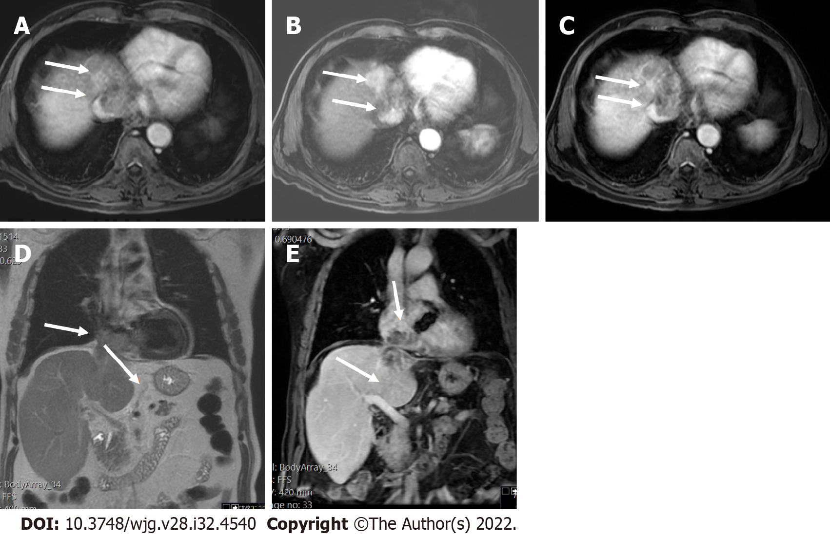
Figure 8 Two-dimensional transthoracic echocardiogram revealed a mobile right atrial mass protruding from the inferior vena cava, in a patient with chronic hepatitis B and cirrhosis.
A-C: Axial T1 weighted image (T1WI) (A), arterial post-contrast (B) and portal phase (C) showed direct right atrial extension of hepatic segment VIII-IVA tumor through the inferior vena cava, categorized as LR-tumor in vein (arrows); D and E: Coronal T2 weighted image (D) and coronal post-contrast T1WI (E) showed better craniocaudal tumor extension (arrows).
- Citation: Liava C, Sinakos E, Papadopoulou E, Giannakopoulou L, Potsi S, Moumtzouoglou A, Chatziioannou A, Stergioulas L, Kalogeropoulou L, Dedes I, Akriviadis E, Chourmouzi D. Liver Imaging Reporting and Data System criteria for the diagnosis of hepatocellular carcinoma in clinical practice: A pictorial minireview. World J Gastroenterol 2022; 28(32): 4540-4556
- URL: https://www.wjgnet.com/1007-9327/full/v28/i32/4540.htm
- DOI: https://dx.doi.org/10.3748/wjg.v28.i32.4540


