Copyright
©The Author(s) 2022.
World J Gastroenterol. Aug 7, 2022; 28(29): 3994-4006
Published online Aug 7, 2022. doi: 10.3748/wjg.v28.i29.3994
Published online Aug 7, 2022. doi: 10.3748/wjg.v28.i29.3994
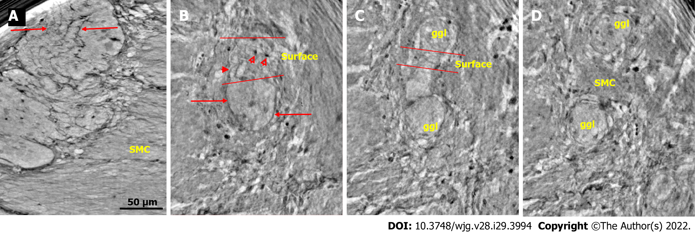
Figure 1 Ganglion from healthy human ileum.
A: Part of the myenteric ganglion in two portions. Quite regular, almost parallel, thin cytoplasmic projections of telocytes (telopodes) with short, thin ”spines” on the surface are observed (between arrows). The scale bar (50 μm) was applied to all subfigures; B: A small portion of the ganglion containing two neurons. On the surface of the ganglion (between the two lines), the cellular nuclei of the two telocytes were observed as dark spots (empty arrowheads). Telopodes radiate from their body. Arrows indicate the telopodes around the ganglion. A telopode can be followed toward the surface (arrowhead); C: 16.9 μm deeper from Figure B, two portions appeared with thin telopodes on the surface; D: 10.1 μm deeper from Figure C, SMCs are observed between the two portions. B-D are representative virtual slices from a series covering 27.0 μm thickness of the ganglion. Ggl: Ganglion; SMC: Smooth muscle cells.
- Citation: Veress B, Peruzzi N, Eckermann M, Frohn J, Salditt T, Bech M, Ohlsson B. Structure of the myenteric plexus in normal and diseased human ileum analyzed by X-ray virtual histology slices. World J Gastroenterol 2022; 28(29): 3994-4006
- URL: https://www.wjgnet.com/1007-9327/full/v28/i29/3994.htm
- DOI: https://dx.doi.org/10.3748/wjg.v28.i29.3994


