Copyright
©The Author(s) 2022.
World J Gastroenterol. Jul 21, 2022; 28(27): 3334-3345
Published online Jul 21, 2022. doi: 10.3748/wjg.v28.i27.3334
Published online Jul 21, 2022. doi: 10.3748/wjg.v28.i27.3334
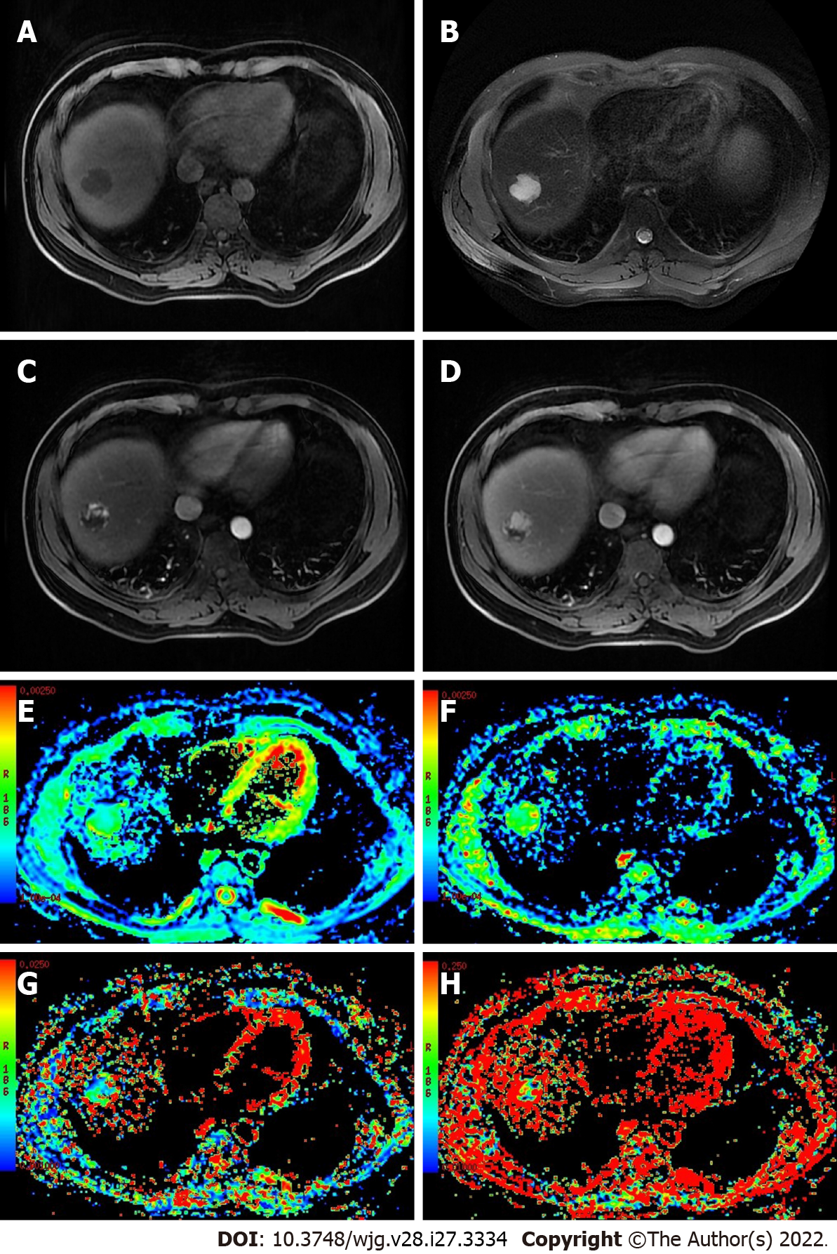
Figure 2 Hepatic hemangiomas in a 35-year-old male.
A 3.0-cm-sized mass in the right hepatic section shows hypointensity on the unenhanced T1-weighted image, hyperintensity on the unenhanced T2-weighted image, peripheral globular enhancement in the image of the arterial phase and centripetal fill-in in the image of the portal venous phase. A: Unenhanced T1-weighted image; B: Unenhanced T2-weighted image; C: Image of the arterial phase; D: Image of the portal venous phase; E: Image showing the apparent diffusion coefficient; F: Image showing the D; G: Image showing the D*; H: Image showing the f.
- Citation: Zhou Y, Zheng J, Yang C, Peng J, Liu N, Yang L, Zhang XM. Application of intravoxel incoherent motion diffusion-weighted imaging in hepatocellular carcinoma. World J Gastroenterol 2022; 28(27): 3334-3345
- URL: https://www.wjgnet.com/1007-9327/full/v28/i27/3334.htm
- DOI: https://dx.doi.org/10.3748/wjg.v28.i27.3334


