Copyright
©The Author(s) 2022.
World J Gastroenterol. Jul 14, 2022; 28(26): 3008-3026
Published online Jul 14, 2022. doi: 10.3748/wjg.v28.i26.3008
Published online Jul 14, 2022. doi: 10.3748/wjg.v28.i26.3008
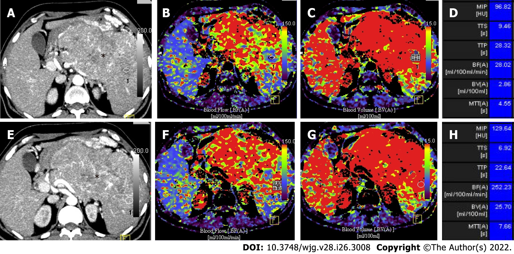
Figure 5 Volume perfusion computed tomography images of a 67-year-old man with a large grade 3 neuroendocrine neoplasm involving body and tail of pancreas.
A and B: Axial arterial phase computed tomography images with circular regions of interest placed at two different locations in the lesion (*); C-H: Parametric maps for blood flow (C and D) and blood volume (E and F) with mean value of each perfusion parameter (G and H) are shown. Lower values of mean blood flow, mean blood volume and mean transit time are features of high grade neuroendocrine neoplasm.
- Citation: Ramachandran A, Madhusudhan KS. Advances in the imaging of gastroenteropancreatic neuroendocrine neoplasms. World J Gastroenterol 2022; 28(26): 3008-3026
- URL: https://www.wjgnet.com/1007-9327/full/v28/i26/3008.htm
- DOI: https://dx.doi.org/10.3748/wjg.v28.i26.3008


