Copyright
©The Author(s) 2022.
World J Gastroenterol. Jul 7, 2022; 28(25): 2843-2853
Published online Jul 7, 2022. doi: 10.3748/wjg.v28.i25.2843
Published online Jul 7, 2022. doi: 10.3748/wjg.v28.i25.2843
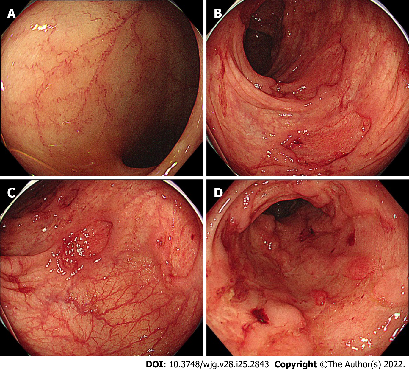
Figure 3 Colonoscopy showing representative endoscopic findings of a patient with Takayasu arteritis and inflammatory bowel disease.
A: Terminal ileum with normal mucosa; B and C: Sigmoid and rectosigmoid colon, respectively, with discontinuous round ulcers; D: Lower rectum, with deep and large longitudinal ulcers.
- Citation: Akiyama S, Fukuda S, Steinberg JM, Suzuki H, Tsuchiya K. Characteristics of inflammatory bowel diseases in patients with concurrent immune-mediated inflammatory diseases. World J Gastroenterol 2022; 28(25): 2843-2853
- URL: https://www.wjgnet.com/1007-9327/full/v28/i25/2843.htm
- DOI: https://dx.doi.org/10.3748/wjg.v28.i25.2843


