Copyright
©The Author(s) 2022.
World J Gastroenterol. Jun 28, 2022; 28(24): 2748-2757
Published online Jun 28, 2022. doi: 10.3748/wjg.v28.i24.2748
Published online Jun 28, 2022. doi: 10.3748/wjg.v28.i24.2748
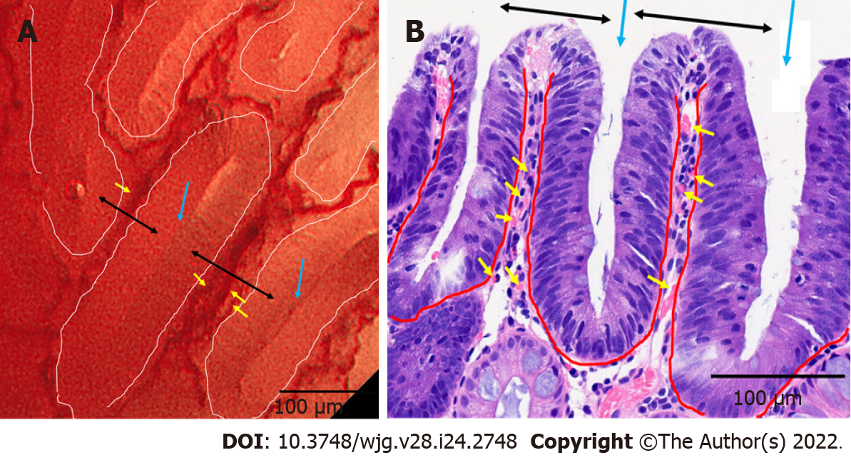
Figure 3 Correspondence between endocytoscopy and histology in a low-grade tubular adenoma.
A: A set of an endocytoscope (CF-H290EC), X1 system, and 4K 32-inch monitor was used. Full-zoom magnifying, narrow band imaging observation. Variably shaped tubular glands (surrounded by white curves) with homogeneous widths of about 100 µm. The crypts (blue arrows) are detected as brown and white slits inside and along the tubular glands. The microvessels (yellow arrows) are identified in the intervening part (black arrows); B: Vertical sections stained with hematoxylin-eosin. The tubular glands (surrounded by red curves), crypts (blue arrows), intervening parts (black arrows), and microvessels (yellow arrows) are seen in endocytoscopy.
- Citation: Toyoshima O, Nishizawa T, Yoshida S, Watanabe H, Odawara N, Sakitani K, Arano T, Takiyama H, Kobayashi H, Kogure H, Fujishiro M. Brown slits for colorectal adenoma crypts on conventional magnifying endoscopy with narrow band imaging using the X1 system. World J Gastroenterol 2022; 28(24): 2748-2757
- URL: https://www.wjgnet.com/1007-9327/full/v28/i24/2748.htm
- DOI: https://dx.doi.org/10.3748/wjg.v28.i24.2748


