Copyright
©The Author(s) 2022.
World J Gastroenterol. Jun 28, 2022; 28(24): 2748-2757
Published online Jun 28, 2022. doi: 10.3748/wjg.v28.i24.2748
Published online Jun 28, 2022. doi: 10.3748/wjg.v28.i24.2748
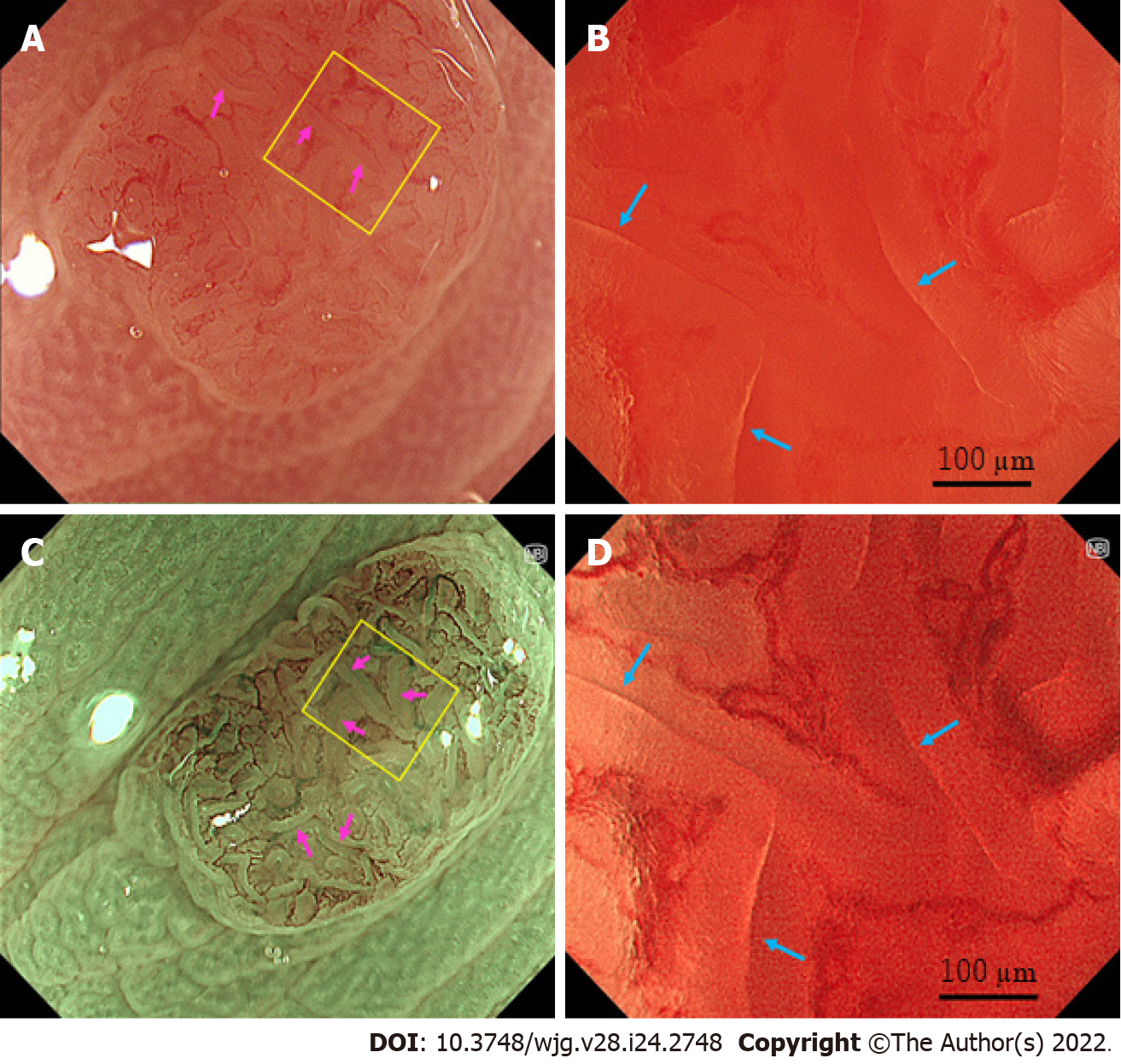
Figure 2 Representative images of “slit-like lumens” and “brown slits” in a low-grade tubular adenoma using endocytoscopy.
A set of an endocytoscope (CF-H290EC), X1 system, and 4K 32-inch monitor was used. A, B: White light imaging observation; C, D: Narrow band imaging observation; A, C: Conventional (100 ×) magnifying observation. The yellow box represents the site for full-zoom observation; B, D: Full-zoom (790 ×) magnifying observation; D: Slit-like lumens were observed (blue arrows). The microvessels were clearly observed surrounding the slit-like lumen and showed a vessel network; C: A “brown slit” sign was observed (pink arrows); B: Slit-like lumens were obscurely observed (blue arrows).
- Citation: Toyoshima O, Nishizawa T, Yoshida S, Watanabe H, Odawara N, Sakitani K, Arano T, Takiyama H, Kobayashi H, Kogure H, Fujishiro M. Brown slits for colorectal adenoma crypts on conventional magnifying endoscopy with narrow band imaging using the X1 system. World J Gastroenterol 2022; 28(24): 2748-2757
- URL: https://www.wjgnet.com/1007-9327/full/v28/i24/2748.htm
- DOI: https://dx.doi.org/10.3748/wjg.v28.i24.2748


