Copyright
©The Author(s) 2022.
World J Gastroenterol. Jun 28, 2022; 28(24): 2733-2747
Published online Jun 28, 2022. doi: 10.3748/wjg.v28.i24.2733
Published online Jun 28, 2022. doi: 10.3748/wjg.v28.i24.2733
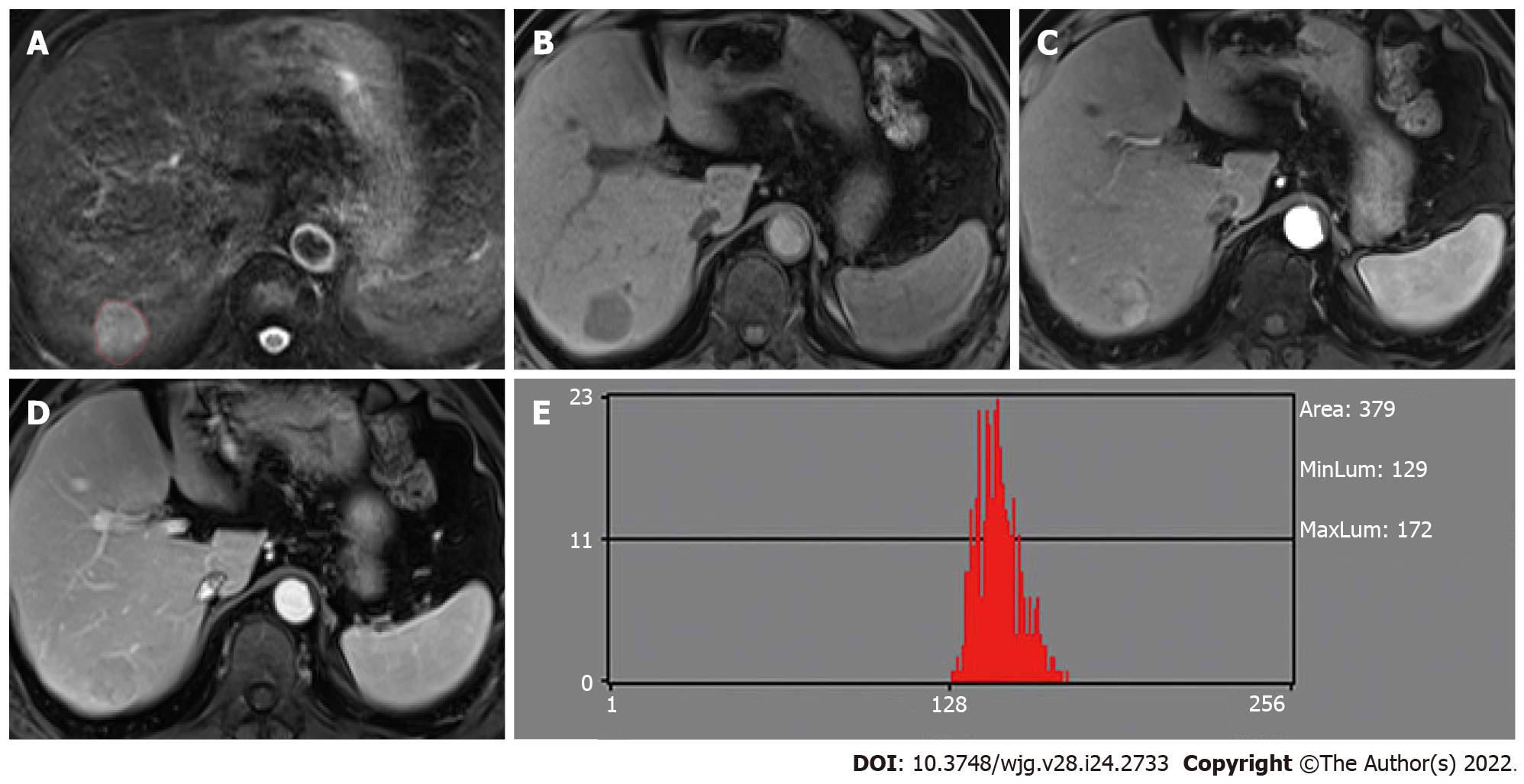
Figure 2 Hepatocellular carcinoma without microvascular invasion in a 60-year-old man.
A: The lesion showed slightly high signal intensity on T2-weighted imaging (T2WI) and was first regions of interest segmented; B: T1WI showed hypointensity; C: Hyper-enhancement in the arterial phase; D: The lesion showed wash-out in the portal venous phase; E: Histogram map derived from the portal venous phase.
- Citation: Li YM, Zhu YM, Gao LM, Han ZW, Chen XJ, Yan C, Ye RP, Cao DR. Radiomic analysis based on multi-phase magnetic resonance imaging to predict preoperatively microvascular invasion in hepatocellular carcinoma. World J Gastroenterol 2022; 28(24): 2733-2747
- URL: https://www.wjgnet.com/1007-9327/full/v28/i24/2733.htm
- DOI: https://dx.doi.org/10.3748/wjg.v28.i24.2733


