Copyright
©The Author(s) 2022.
World J Gastroenterol. Jun 28, 2022; 28(24): 2654-2666
Published online Jun 28, 2022. doi: 10.3748/wjg.v28.i24.2654
Published online Jun 28, 2022. doi: 10.3748/wjg.v28.i24.2654
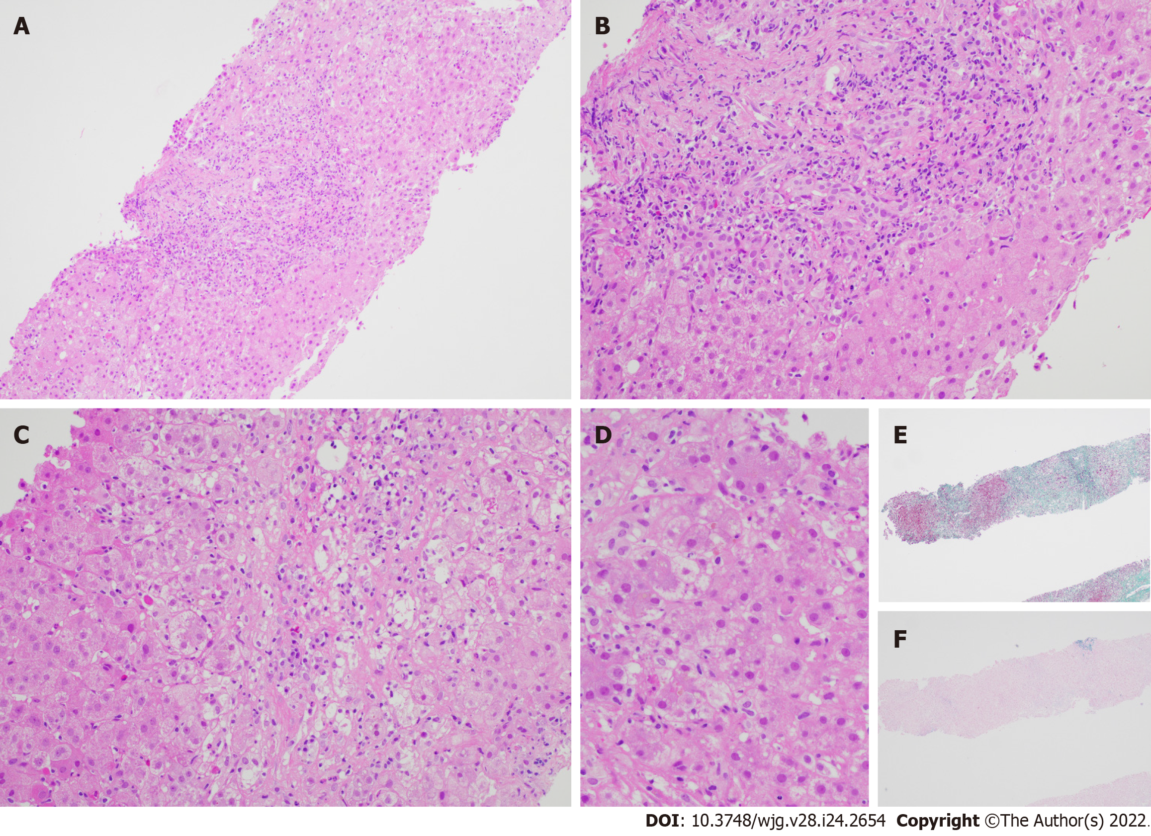
Figure 1 Liver biopsy specimen for patient A.
A: Low power view [hematoxylin & eosin (H&E) 100 ×] displays conspicuous portal and lobular inflammation with lobular disarray. Mild steatosis is also noted; B: Higher magnification of the portal tract (H&E 200 ×), zone 1, shows moderate chronic inflammation, lymphoplasmacytic predominantly, and rare eosinophils, with interface damage; C: At similar magnification (H&E 200 ×), the lobule including the perivenular region, e.g., zones 2 and 3, exhibits lobulitis characterized by aggregates of plasma cells, swollen hepatocytes with rosetting, Councilman bodies, and hepatocyte drop-out; D: High power view (H&E 400 ×) demonstrates rosetting of hepatocytes with droplets of orange-brown bile pigment; E and F: Histochemical stains Masson trichrome (E, 40 ×) showing collapse with mild early young fibrosis and Victoria blue (F, 40 ×) revealing paucity of elastic fibers, thus in keeping with subacute injury. Overall, the appearances are supportive of subacute drug-induced liver injury in association with autoimmune hepatitis histological pattern.
- Citation: Tan CK, Ho D, Wang LM, Kumar R. Drug-induced autoimmune hepatitis: A minireview. World J Gastroenterol 2022; 28(24): 2654-2666
- URL: https://www.wjgnet.com/1007-9327/full/v28/i24/2654.htm
- DOI: https://dx.doi.org/10.3748/wjg.v28.i24.2654


