Copyright
©The Author(s) 2022.
World J Gastroenterol. Jun 21, 2022; 28(23): 2625-2632
Published online Jun 21, 2022. doi: 10.3748/wjg.v28.i23.2625
Published online Jun 21, 2022. doi: 10.3748/wjg.v28.i23.2625
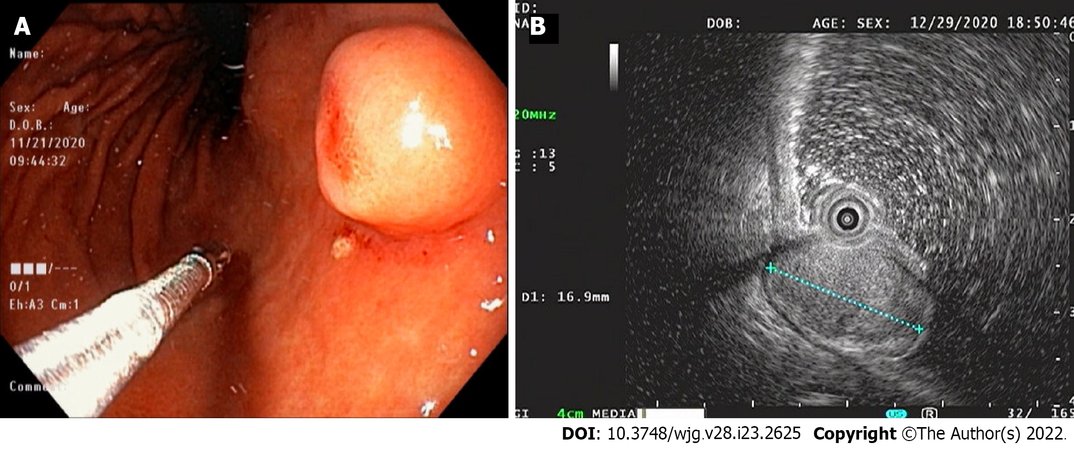
Figure 1 Endoscopy and endoscopic ultrasound images.
A: Endoscopic image showing a 15-mm-sized, submucosal tumor-like, protruding lesion with focal mucosal erythema and depression of overlying mucosa; B: Endoscopic ultrasound image showing a well-circumscribed, slightly heterogeneous, 17 mm × 10 mm sized, isoechoic mass originating from the third sonographic layer.
- Citation: Cho JH, Byeon JH, Lee SH. Primary gastric dedifferentiated liposarcoma resected endoscopically: A case report. World J Gastroenterol 2022; 28(23): 2625-2632
- URL: https://www.wjgnet.com/1007-9327/full/v28/i23/2625.htm
- DOI: https://dx.doi.org/10.3748/wjg.v28.i23.2625


