Copyright
©The Author(s) 2022.
World J Gastroenterol. May 7, 2022; 28(17): 1751-1767
Published online May 7, 2022. doi: 10.3748/wjg.v28.i17.1751
Published online May 7, 2022. doi: 10.3748/wjg.v28.i17.1751
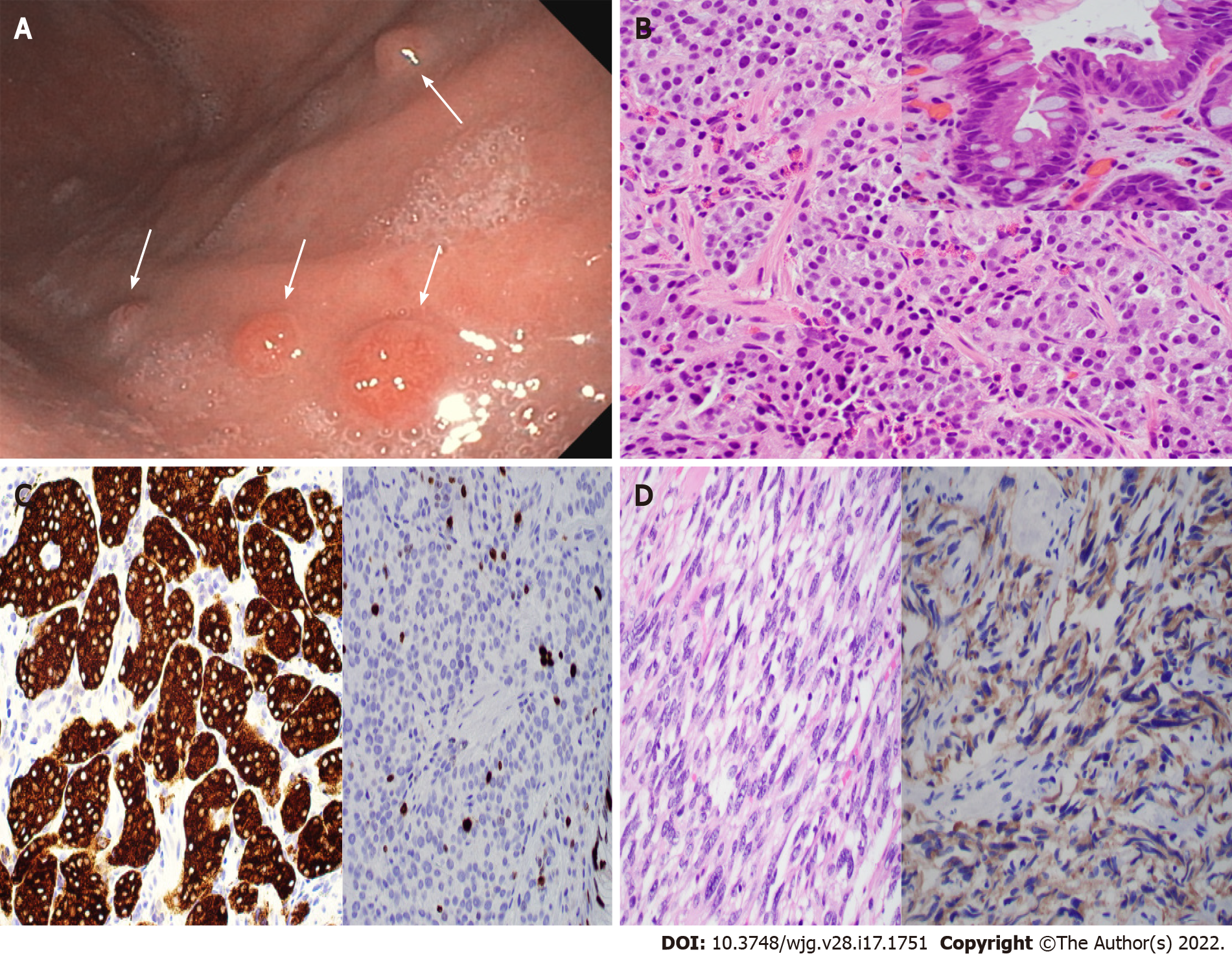
Figure 2 Type 1 gastric neuroendocrine tumor with usual and unusual histology.
A: Endoscopic photo showing four nodules (arrows) in the gastric body of a 56-year-old woman with a history of autoimmune chronic atrophic gastritis (ACAG); B and C: Biopsy of the nodules showing well-differentiated neuroendocrine tumor (B, H&E stain, 400 ×) with intestinal metaplasia on the overlying mucosa (B inset), positivity for synaptophysin (C, left), and a 7% Ki67 index (C, right); D: Gastric biopsy in an 83-year-old woman with ACAG showing a neuroendocrine tumor with unusual spindle cell morphology mimicking gastrointestinal stromal tumor (GIST) (left, H&E stain), which is positive for synaptophysin (right) and negative for GIST markers.
- Citation: Yin F, Wu ZH, Lai JP. New insights in diagnosis and treatment of gastroenteropancreatic neuroendocrine neoplasms. World J Gastroenterol 2022; 28(17): 1751-1767
- URL: https://www.wjgnet.com/1007-9327/full/v28/i17/1751.htm
- DOI: https://dx.doi.org/10.3748/wjg.v28.i17.1751


