Copyright
©The Author(s) 2022.
World J Gastroenterol. Apr 7, 2022; 28(13): 1315-1328
Published online Apr 7, 2022. doi: 10.3748/wjg.v28.i13.1315
Published online Apr 7, 2022. doi: 10.3748/wjg.v28.i13.1315
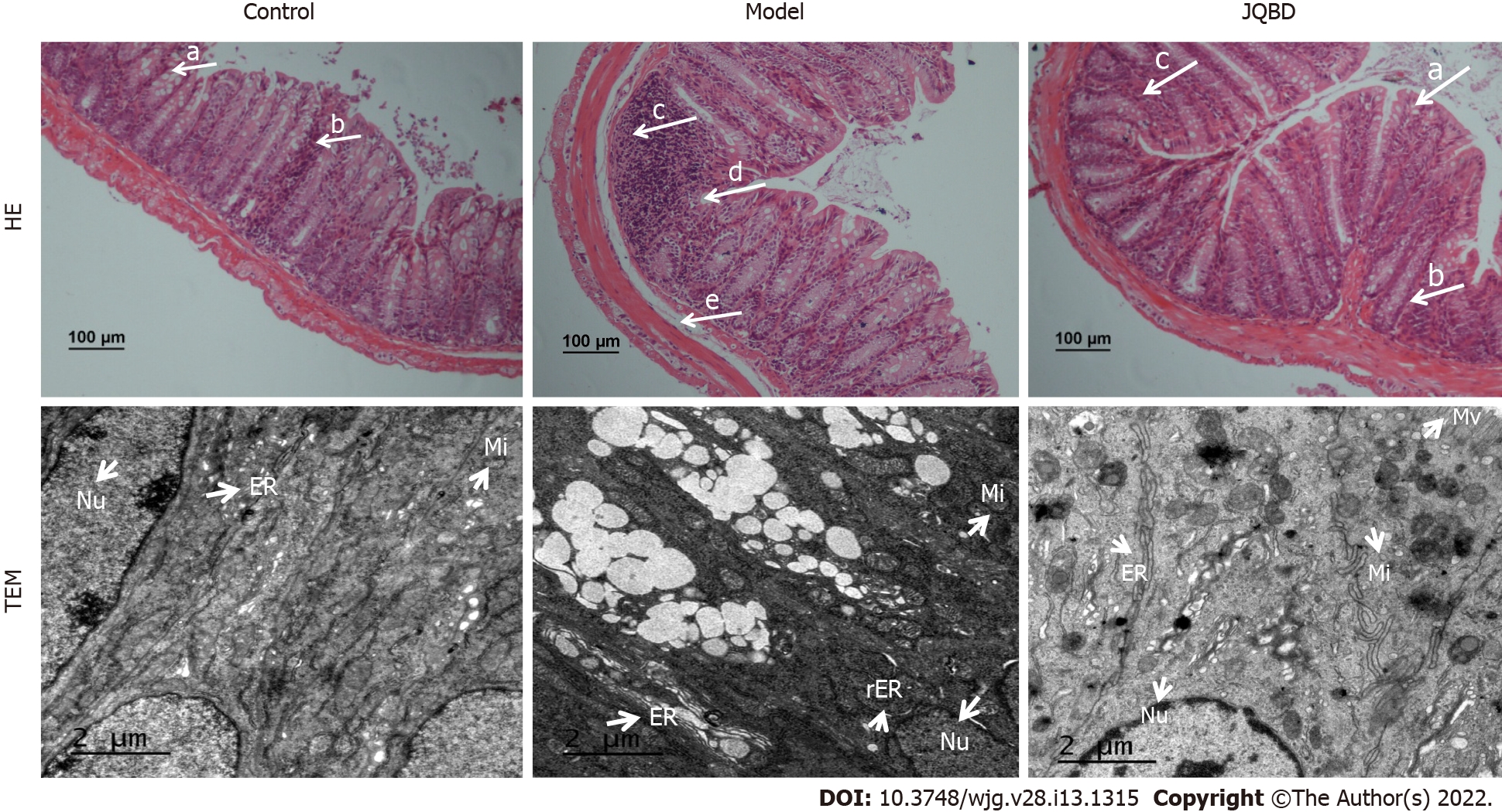
Figure 4 Histological evaluation of the colonic mucosa following hematoxylin and eosin staining (× 100) and ultrastructure of the colonic epithelium by transmission electron microscopy (× 6000).
Arrows indicate goblet cells (a), crypts (b), inflammatory cells infiltration (c), epithelium surface erosion (d), and submucosal oedema (e). Control: Control group; Model: Model group; JQBD: Jianpi Qingchang Bushen decoction Group; Nu: Nucleus; Mi: Mitochondrial; ER: Endoplasmic Reticulum; rER: Rough endoplasmic reticulum; Mv: Microvillus.
- Citation: Zhang YL, Chen Q, Zheng L, Zhang ZW, Chen YJ, Dai YC, Tang ZP. Jianpi Qingchang Bushen decoction improves inflammatory response and metabolic bone disorder in inflammatory bowel disease-induced bone loss. World J Gastroenterol 2022; 28(13): 1315-1328
- URL: https://www.wjgnet.com/1007-9327/full/v28/i13/1315.htm
- DOI: https://dx.doi.org/10.3748/wjg.v28.i13.1315


