Copyright
©The Author(s) 2022.
World J Gastroenterol. Mar 14, 2022; 28(10): 976-984
Published online Mar 14, 2022. doi: 10.3748/wjg.v28.i10.976
Published online Mar 14, 2022. doi: 10.3748/wjg.v28.i10.976
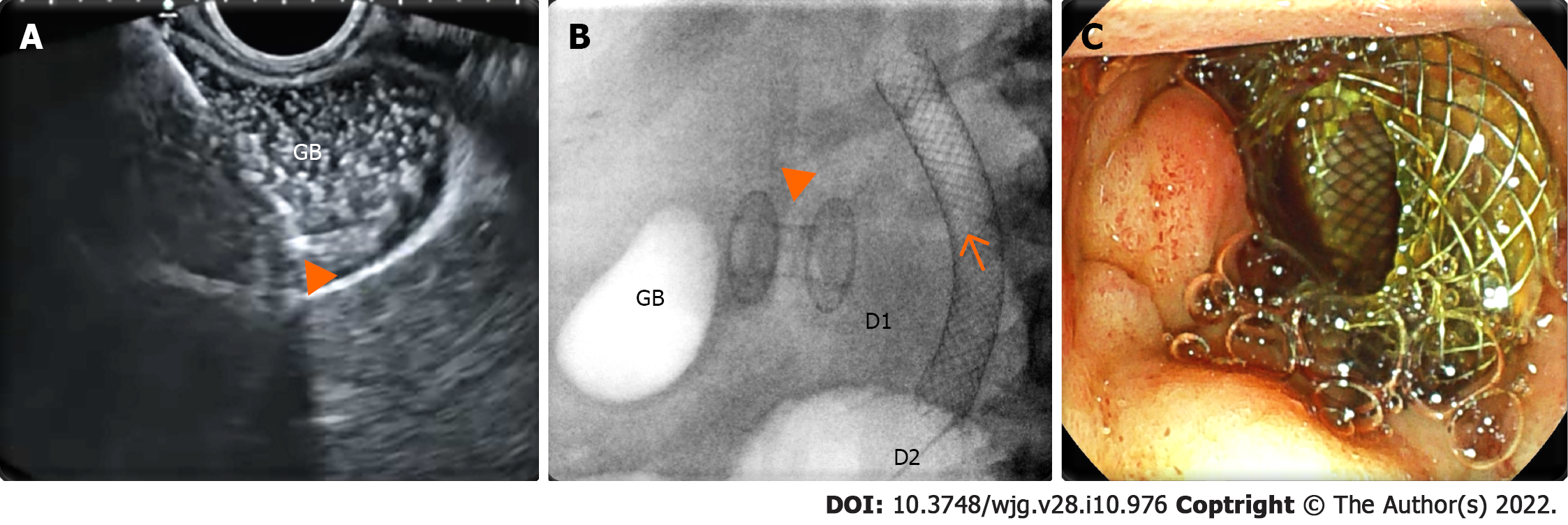
Figure 3 Endoscopic ultrasound-guided gallbladder drainage.
A: Endosonographic view of the delivery system (arrowhead) of the enhanced lumen apposing metal stent inside a distended gallbladder (GB) full of sludge; B: Radioscopic view of the lumen apposing metal stent (arrowhead) released between the gallbladder and duodenal bulb [D1; biliary stent (arrow) released in the second duodenal portion (D2)]; C: Endoscopic view of the proximal flange of the lumen apposing metal stent inside the duodenal bulb.
- Citation: Vanella G, Tamburrino D, Capurso G, Bronswijk M, Reni M, Dell'Anna G, Crippa S, Van der Merwe S, Falconi M, Arcidiacono PG. Feasibility of therapeutic endoscopic ultrasound in the bridge-to-surgery scenario: The example of pancreatic adenocarcinoma. World J Gastroenterol 2022; 28(10): 976-984
- URL: https://www.wjgnet.com/1007-9327/full/v28/i10/976.htm
- DOI: https://dx.doi.org/10.3748/wjg.v28.i10.976


