Copyright
©The Author(s) 2021.
World J Gastroenterol. Dec 14, 2021; 27(46): 7894-7908
Published online Dec 14, 2021. doi: 10.3748/wjg.v27.i46.7894
Published online Dec 14, 2021. doi: 10.3748/wjg.v27.i46.7894
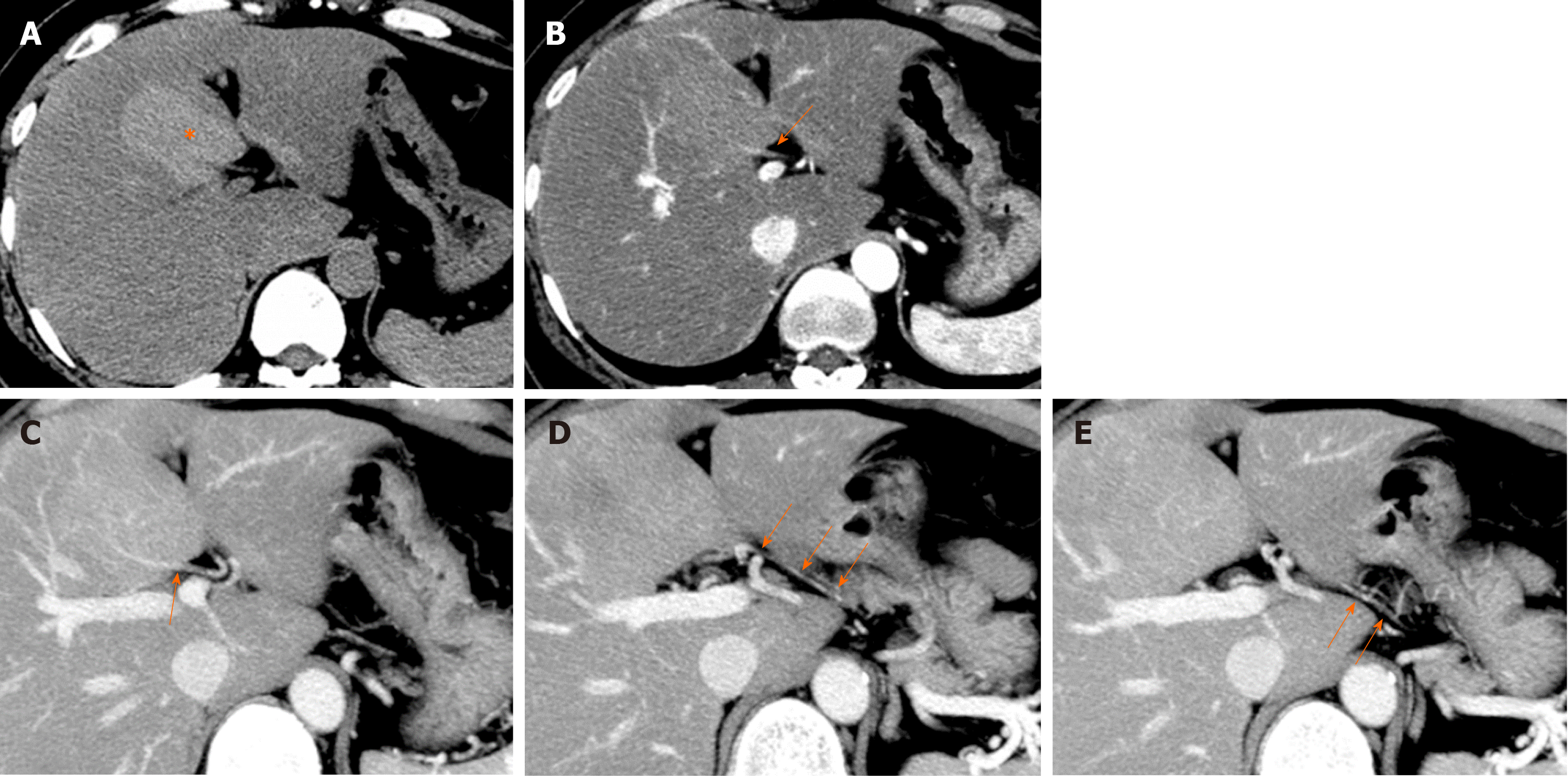
Figure 15 Focal spared area of fatty liver in posterior aspect of segment IV (40th female).
A: On pre-contrast enhanced computed tomography (CT), a focal hyperdense lesion compared to the background liver parenchyma is observed in posterior aspect of segment IV of the liver (*). Background liver shows decreased density and suggestive of fatty liver and hyperdese area is diagnosed as focal sparing of fatty liver. B: On arterial phase contrast enhanced CT image, an enhanced vascular branch is directly entering the area at hepatic hilum (arrow). C-E: On sequential images of arterial phase contrast enhanced CT, aberrant right gastric vein directly entering to the posterior aspect of segment IV of the liver is well opacified (arrows). These findings represent focal spared area of the fatty liver in aberrant right gastric venous drainage area of the liver at the posterior aspect of segment IV of the liver.
- Citation: Kobayashi S. Hepatic pseudolesions caused by alterations in intrahepatic hemodynamics. World J Gastroenterol 2021; 27(46): 7894-7908
- URL: https://www.wjgnet.com/1007-9327/full/v27/i46/7894.htm
- DOI: https://dx.doi.org/10.3748/wjg.v27.i46.7894


