Copyright
©The Author(s) 2021.
World J Gastroenterol. Dec 14, 2021; 27(46): 7866-7893
Published online Dec 14, 2021. doi: 10.3748/wjg.v27.i46.7866
Published online Dec 14, 2021. doi: 10.3748/wjg.v27.i46.7866
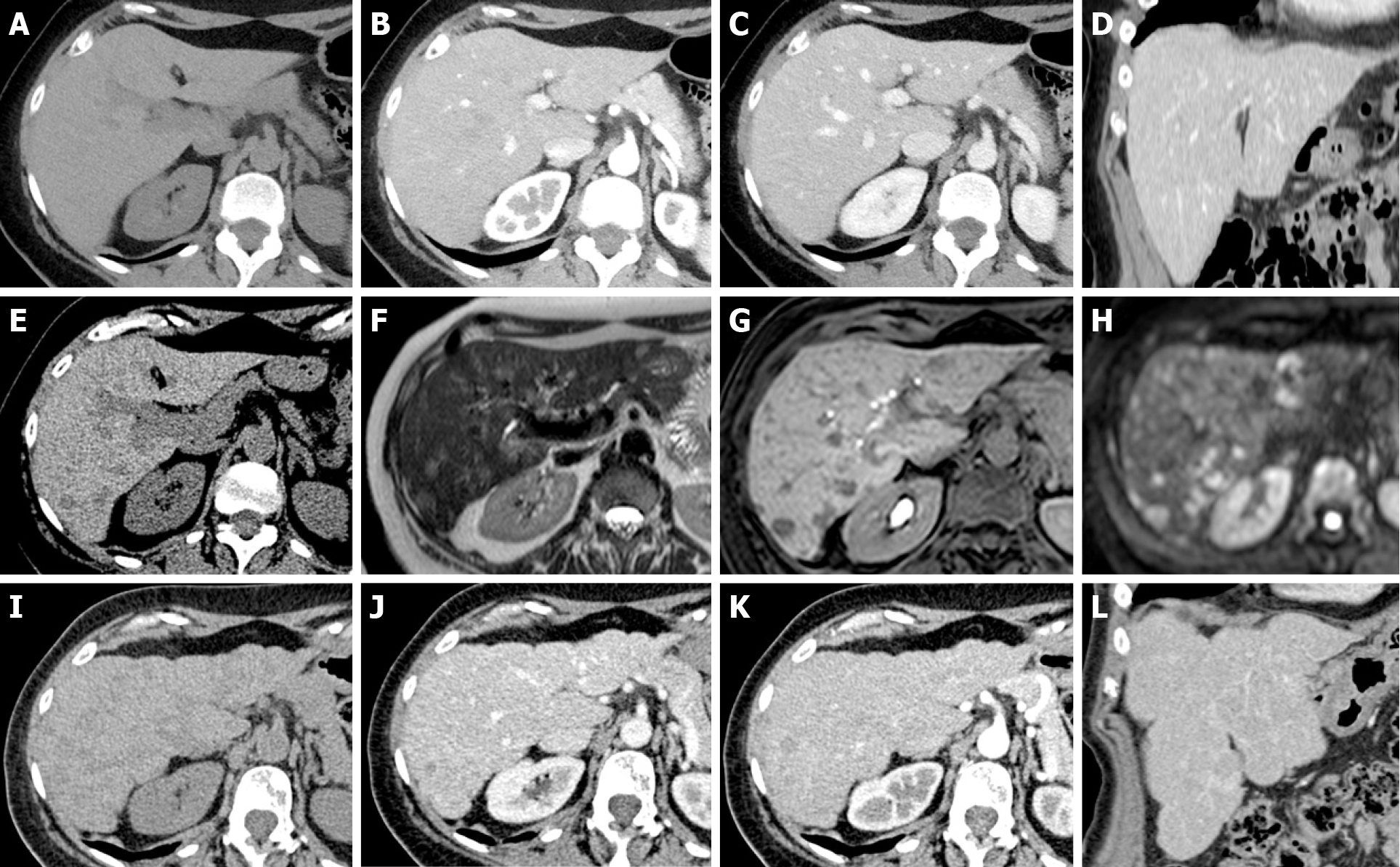
Figure 15 Pseudocirrhosis in patients with breast cancer treated with surgery and 6 mo of chemotherapy (capecitabine and monoclonal antibodies).
A-D: Unenhanced (A: Axial) and contrast-enhanced computed tomography (CT) (B: Axial arterial phase; C: Axial portal phase; D: Coronal portal phase) was performed at staging. The liver shows regular volume, morphology and a smooth surface. No focal lesions were found; thus, no chemotherapy was undertaken; E: At the 1-year follow-up, unenhanced CT demonstrated the appearance of a hypodense focal lesion (arrowhead); F-H: A complete magnetic resonance study with liver-specific contrast agent confirmed the presence of new focal lesions consistent with metastases. Mild hyperintensity in the T2w sequence (F), clear hypointensity in the fat sat gradient echo 3D T1w hepatobiliary phase (G) and high signal in diffusion-weighted images (H) are shown. Chemotherapy was started. I-L: A 6-mo follow-up unenhanced (I: Axial) and contrast-enhanced CT (J: Axial arterial phase; K: Axial portal phase; L: Coronal portal phase) shows typical signs of liver pseudocirrhotic changes: parenchymal volume reduction, irregular macrocyclic margins, right lobe atrophy and caudate lobe hypertrophy.
- Citation: Calistri L, Rastrelli V, Nardi C, Maraghelli D, Vidali S, Pietragalla M, Colagrande S. Imaging of the chemotherapy-induced hepatic damage: Yellow liver, blue liver, and pseudocirrhosis. World J Gastroenterol 2021; 27(46): 7866-7893
- URL: https://www.wjgnet.com/1007-9327/full/v27/i46/7866.htm
- DOI: https://dx.doi.org/10.3748/wjg.v27.i46.7866


