Copyright
©The Author(s) 2021.
World J Gastroenterol. Dec 14, 2021; 27(46): 7866-7893
Published online Dec 14, 2021. doi: 10.3748/wjg.v27.i46.7866
Published online Dec 14, 2021. doi: 10.3748/wjg.v27.i46.7866
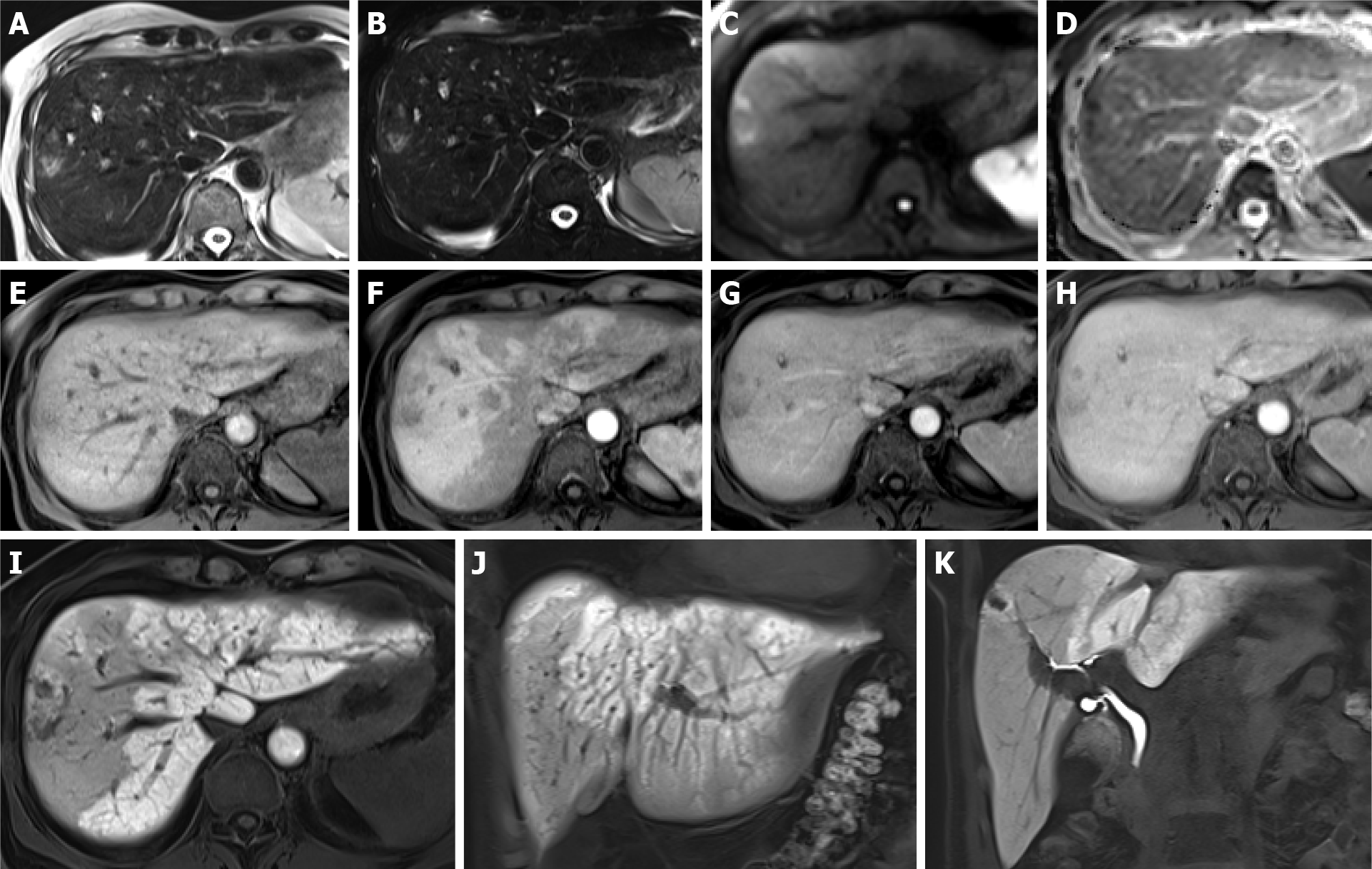
Figure 6 Association between sinusoidal obstructive syndrome and peliosis in patients without a history of hepatopathy, with lung cancer treated with 3 cycles of cisplatin and etoposide and 12 subsequent cycles of immunotherapy.
A-K: After 1 year of therapy, magnetic resonance was performed for abdominal pain and an increase in liver enzymes. Hyperintense nodules on T2w (A), T2w fat sat (B) and high b-value diffusion-weighted images (C) are seen. On the apparent diffusion coefficient map (D), they are hypointense. On dynamic imaging, weak enhancement is seen (E: Unenhanced image; F, G, H: Arterial, portal and equilibrium phases, respectively). In the hepatobiliary phase, they appear predominantly hypointense (I-K). A transcutaneous biopsy was performed, resulting in peliosis nodules. Mosaic pattern enhancement of the liver parenchyma in the arterial phase (F) and reticular aspects in the hepatobiliary phase (I-J) were consistent with sinusoidal obstructive syndrome.
- Citation: Calistri L, Rastrelli V, Nardi C, Maraghelli D, Vidali S, Pietragalla M, Colagrande S. Imaging of the chemotherapy-induced hepatic damage: Yellow liver, blue liver, and pseudocirrhosis. World J Gastroenterol 2021; 27(46): 7866-7893
- URL: https://www.wjgnet.com/1007-9327/full/v27/i46/7866.htm
- DOI: https://dx.doi.org/10.3748/wjg.v27.i46.7866


