Copyright
©The Author(s) 2021.
World J Gastroenterol. Dec 14, 2021; 27(46): 7866-7893
Published online Dec 14, 2021. doi: 10.3748/wjg.v27.i46.7866
Published online Dec 14, 2021. doi: 10.3748/wjg.v27.i46.7866
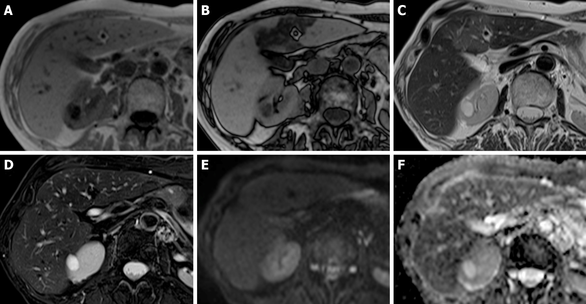
Figure 5 Chemotherapy-associated focal steatosis in patients with lung cancer 3 mo after the beginning of immunotherapy.
A-F: On magnetic resonance, focal geographic fatty deposition, poorly delineated, is seen as signal hypointensity on gradient echo (GE) T1w out-of-phase (B) in the periportal aspect of segment IV and around the falciform ligament. On T2w images (C) it is weakly hyperintense. No signal alterations are seen on the GE T1w in-phase images (A), T2w fat saturation (D), diffusion-weighted (E) images, or apparent diffusion coefficient map (F).
- Citation: Calistri L, Rastrelli V, Nardi C, Maraghelli D, Vidali S, Pietragalla M, Colagrande S. Imaging of the chemotherapy-induced hepatic damage: Yellow liver, blue liver, and pseudocirrhosis. World J Gastroenterol 2021; 27(46): 7866-7893
- URL: https://www.wjgnet.com/1007-9327/full/v27/i46/7866.htm
- DOI: https://dx.doi.org/10.3748/wjg.v27.i46.7866


