Copyright
©The Author(s) 2021.
World J Gastroenterol. Oct 14, 2021; 27(38): 6501-6510
Published online Oct 14, 2021. doi: 10.3748/wjg.v27.i38.6501
Published online Oct 14, 2021. doi: 10.3748/wjg.v27.i38.6501
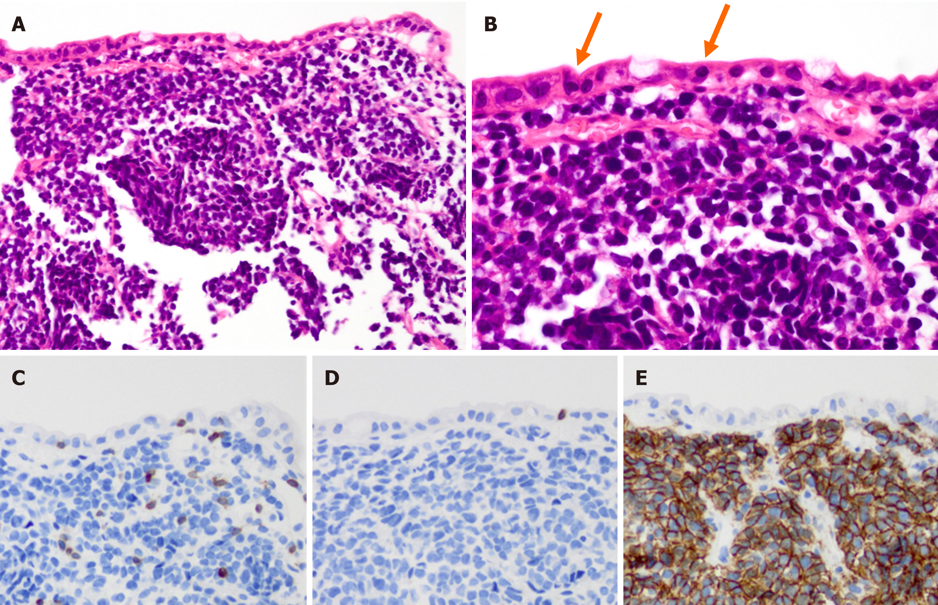
Figure 2 Histological and immunohistochemical analyses.
A: Hematoxylin-eosin staining showed infiltration of medium-size atypical lymphoid cells in the mucosa; B: Lymphoid cell infiltration was also observed in the epithelium (arrow); C: Lymphoid cells expressed CD3; D: The lymphoid cells were CD8 negative; E: Lymphoid cells were CD56 positive. Magnification 200× (A, C-E) and 400× (B).
- Citation: Ozaka S, Inoue K, Okajima T, Tasaki T, Ariki S, Ono H, Ando T, Daa T, Murakami K. Monomorphic epitheliotropic intestinal T-cell lymphoma presenting as melena with long-term survival: A case report and review of literature. World J Gastroenterol 2021; 27(38): 6501-6510
- URL: https://www.wjgnet.com/1007-9327/full/v27/i38/6501.htm
- DOI: https://dx.doi.org/10.3748/wjg.v27.i38.6501


