Copyright
©The Author(s) 2021.
World J Gastroenterol. Oct 14, 2021; 27(38): 6501-6510
Published online Oct 14, 2021. doi: 10.3748/wjg.v27.i38.6501
Published online Oct 14, 2021. doi: 10.3748/wjg.v27.i38.6501
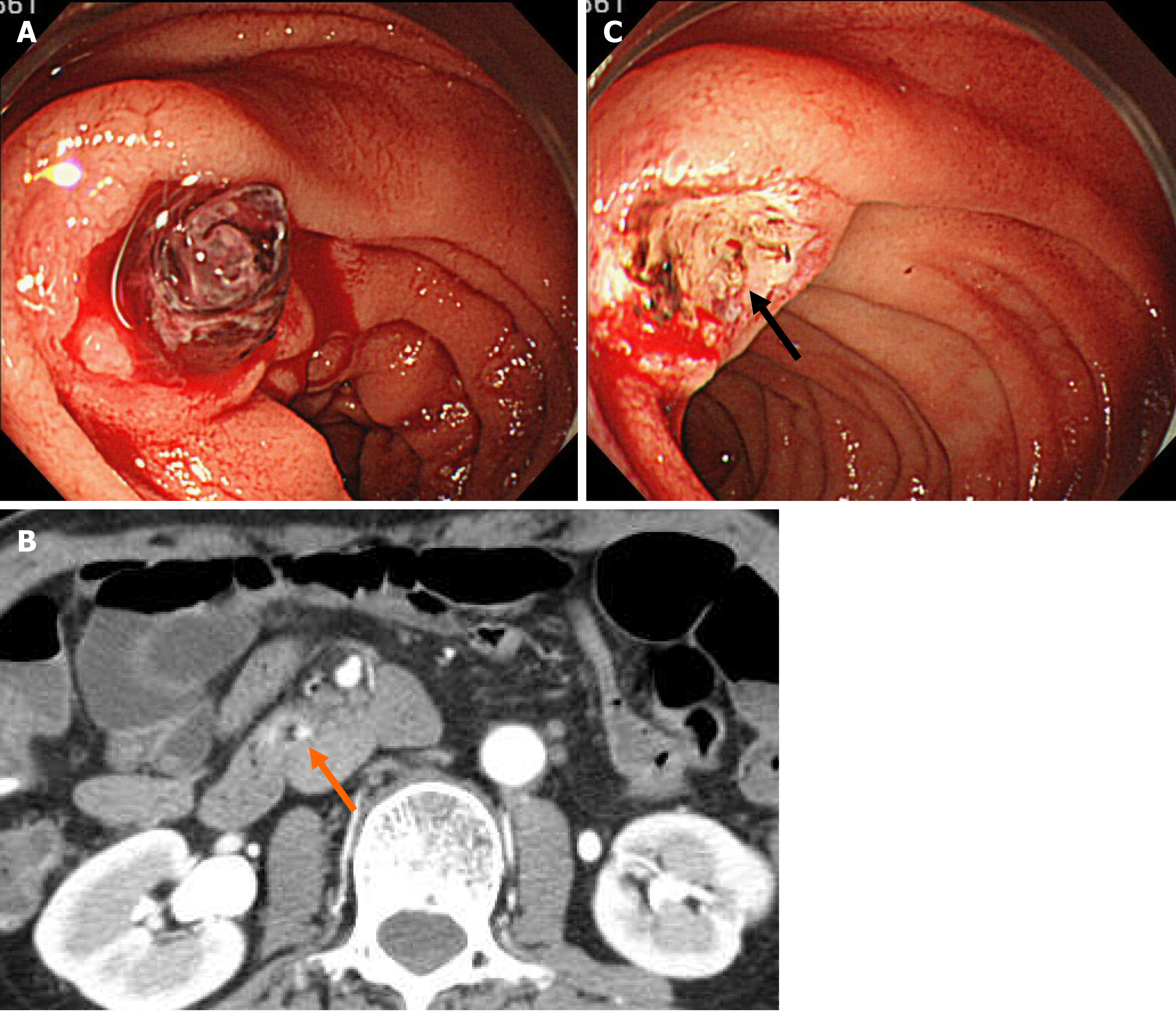
Figure 1 Endoscopic images and abdominal contrast-enhanced computed tomography scan images on initial examination.
A: Esophagogastroduodenoscopy showed an ulcerative lesion with fresh blood clots in the transverse part of the duodenum; B: Contrast-enhanced computed tomography scan showed a slight localized contrast effect on the wall of the transverse part of the duodenum (arrow); C: When the blood clots were removed, a protruding vessel was observed at the base of the ulcer (arrow).
- Citation: Ozaka S, Inoue K, Okajima T, Tasaki T, Ariki S, Ono H, Ando T, Daa T, Murakami K. Monomorphic epitheliotropic intestinal T-cell lymphoma presenting as melena with long-term survival: A case report and review of literature. World J Gastroenterol 2021; 27(38): 6501-6510
- URL: https://www.wjgnet.com/1007-9327/full/v27/i38/6501.htm
- DOI: https://dx.doi.org/10.3748/wjg.v27.i38.6501


