Copyright
©The Author(s) 2021.
World J Gastroenterol. Oct 7, 2021; 27(37): 6332-6344
Published online Oct 7, 2021. doi: 10.3748/wjg.v27.i37.6332
Published online Oct 7, 2021. doi: 10.3748/wjg.v27.i37.6332
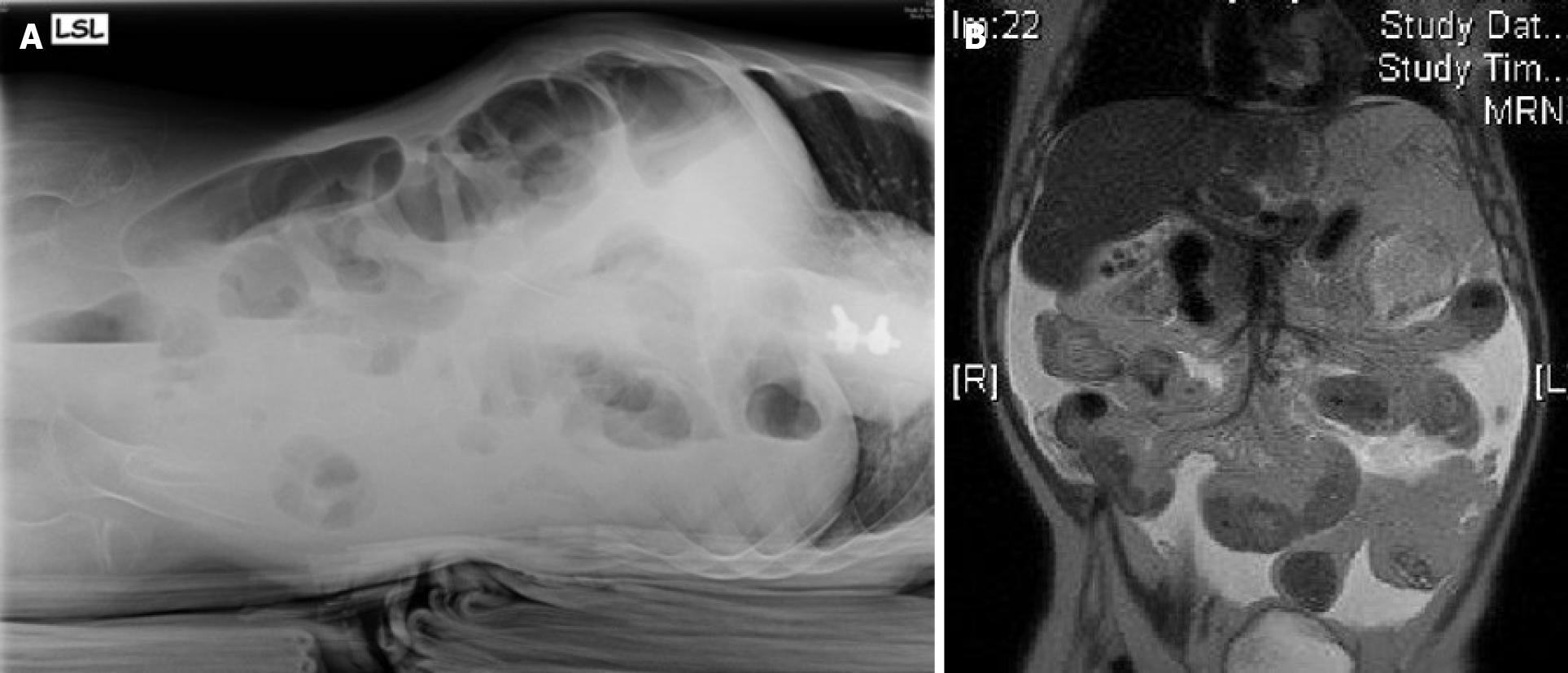
Figure 4 Diagnostic imaging.
A: X-ray with lateral abdominal view showing some air fluid levels; B: Magnetic resonance imaging in coronal plane with distended duodenum and small bowel loops with multiple intraluminal air fluid levels and thickened walls.
- Citation: Keese D, Schmedding A, Saalabian K, Lakshin G, Fiegel H, Rolle U. Abdominal cocoon in children: A case report and review of literature. World J Gastroenterol 2021; 27(37): 6332-6344
- URL: https://www.wjgnet.com/1007-9327/full/v27/i37/6332.htm
- DOI: https://dx.doi.org/10.3748/wjg.v27.i37.6332


