Copyright
©The Author(s) 2021.
World J Gastroenterol. Jul 14, 2021; 27(26): 4208-4220
Published online Jul 14, 2021. doi: 10.3748/wjg.v27.i26.4208
Published online Jul 14, 2021. doi: 10.3748/wjg.v27.i26.4208
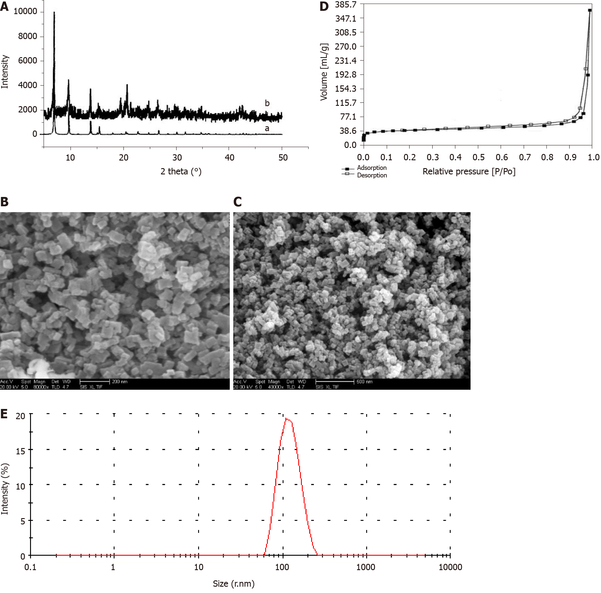
Figure 1 Structure and morphology of IRMOF-3 and norcantharidin-loaded metal-organic framework IRMOF-3 coated with temperature-sensitive gel.
A: X-ray diffraction (XRD) spectra of IRMOF-3. Note: XRD patterns of synthetic IRMOF-3 (b) and standard IRMOF-3 (a). XRD patterns of the sample show the same peaks as those of the standard, confirming the high purity of IRMOF-3. The peak patterns of the sample account for the rough appearance of the nanosized particles; B: Scanning electron microscopy (SEM) images of the morphology of IRMOF-3. SEM images of the morphology of IRMOF-3. The particles show a regular square, uniform distribution and a size of 50-100 nm. The single-particle surface is rough, indicating the existence of pores; C: SEM images of the morphology of norcantharidin (NCTD)-loaded metal-organic framework IRMOF-3 coated with temperature-sensitive gel (NCTD-IRMOF-3-Gel). SEM images of the morphology of NCTD-IRMOF-3-Gel showing the same size as that of IRMOF-3; D: Nitrogen adsorption–desorption isotherms of IRMOF-3. Nitrogen adsorption–desorption isotherms. A hysteresis loop phenomenon appears under relatively high pressure, indicating the existence of channels in the sample; E: Particle size distribution of NCTD-IRMOF-3-Gel. The particle size distribution of NCTD-IRMOF-3-Gel indicated that the average particle size was 100 nm.
- Citation: Li XY, Guan QX, Shang YZ, Wang YH, Lv SW, Yang ZX, Wang R, Feng YF, Li WN, Li YJ. Metal-organic framework IRMOFs coated with a temperature-sensitive gel delivering norcantharidin to treat liver cancer. World J Gastroenterol 2021; 27(26): 4208-4220
- URL: https://www.wjgnet.com/1007-9327/full/v27/i26/4208.htm
- DOI: https://dx.doi.org/10.3748/wjg.v27.i26.4208


