Copyright
©The Author(s) 2021.
World J Gastroenterol. Jan 14, 2021; 27(2): 162-175
Published online Jan 14, 2021. doi: 10.3748/wjg.v27.i2.162
Published online Jan 14, 2021. doi: 10.3748/wjg.v27.i2.162
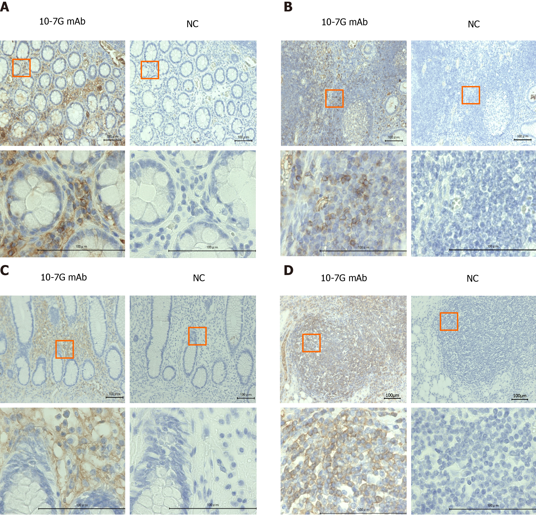
Figure 2 Immunohistochemical study of ulcerative colitis and Crohn’s disease intestinal tissues using the 10-7G mAb.
A and B: Lymphocytes infiltrating into inflammatory sites of the mucosal layer (A) and lymph nodules in intestinal tissues (B) of patients with ulcerative colitis (n = 5) were stained using the 10-7G mAb; C and D: Lymphocytes infiltrating into inflammatory sites of the mucosal layer (C) and lymph nodules in intestinal tissues (D) of patients with Crohn’s disease (n = 5) were also stained. Photographs were acquired using a 20 × (upper) or 100 × (lower) objective. Scale bar, 100 µm. Positive staining was judged by comparison to the negative control. NC: Negative control.
- Citation: Motooka K, Morishita K, Ito N, Shinzaki S, Tashiro T, Nojima S, Shimizu K, Date M, Sakata N, Yamada M, Takamatsu S, Kamada Y, Iijima H, Mizushima T, Morii E, Takehara T, Miyoshi E. Detection of fucosylated haptoglobin using the 10-7G antibody as a biomarker for evaluating endoscopic remission in ulcerative colitis. World J Gastroenterol 2021; 27(2): 162-175
- URL: https://www.wjgnet.com/1007-9327/full/v27/i2/162.htm
- DOI: https://dx.doi.org/10.3748/wjg.v27.i2.162


