Copyright
©The Author(s) 2021.
World J Gastroenterol. May 21, 2021; 27(19): 2299-2311
Published online May 21, 2021. doi: 10.3748/wjg.v27.i19.2299
Published online May 21, 2021. doi: 10.3748/wjg.v27.i19.2299
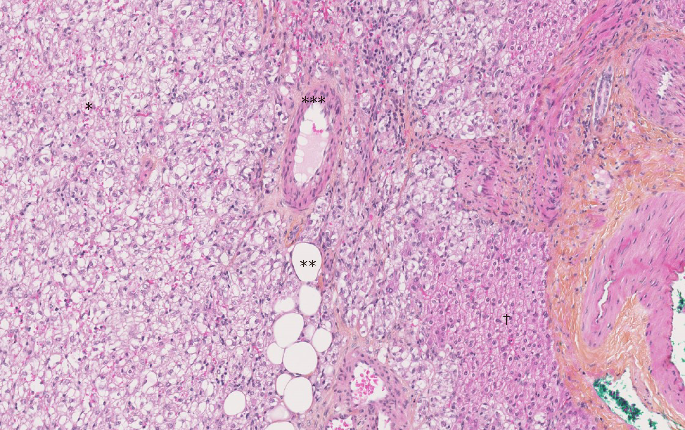
Figure 1 The Hematoxylin-Eosin-Saffron staining image of hepatic angiomyolipoma.
There are three components of hepatic angiomyolipoma: vessel (*), adipocytes (**) and numerous epithelioid cells (***). There are fewer hepatocytes (†) (magnification × 10).
- Citation: Calame P, Tyrode G, Weil Verhoeven D, Félix S, Klompenhouwer AJ, Di Martino V, Delabrousse E, Thévenot T. Clinical characteristics and outcomes of patients with hepatic angiomyolipoma: A literature review. World J Gastroenterol 2021; 27(19): 2299-2311
- URL: https://www.wjgnet.com/1007-9327/full/v27/i19/2299.htm
- DOI: https://dx.doi.org/10.3748/wjg.v27.i19.2299


